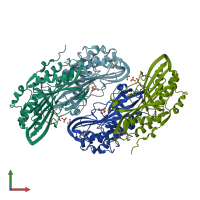
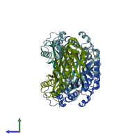
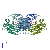
3bp1

X-ray diffraction
1.53Å resolution

Crystal structure of putative 7-cyano-7-deazaguanine reductase QueF from Vibrio cholerae O1 biovar eltor

Released:

Function and Biology Details

Structure analysis Details

Assemblies composition:
homo tetramer
homo dimer (preferred)
PDBe Complex ID:
PDB-CPX-192092 (preferred)
Entry contents:
1 distinct polypeptide molecule
Macromolecule:
NADPH-dependent 7-cyano-7-deazaguanine reductase Chains: A, B, C, D
Molecule details ›
Chains: A, B, C, D
Length: 290 amino acids
Theoretical weight: 33.32 KDa
Source organism: Vibrio cholerae O1 biovar El Tor str. N16961
Expression system: Escherichia coli BL21(DE3)
UniProt:
  • Canonical: Q9KTK0 (Residues: 1-281; Coverage: 100%)
Gene names: VC_0902, queF
Sequence domains:
Structure domains: GTP Cyclohydrolase I, domain 2

Ligands and Environments

1 modified residue:

Experiments and Validation Details

Entry percentile scores
X-ray source: APS BEAMLINE 19-BM
Spacegroup: P1
Unit cell:
a: 71.517Å b: 71.578Å c: 71.511Å
α: 119.25° β: 110.18° γ: 99.58°
R-values:
R R work R free
0.145 0.143 0.183
Expression system: Escherichia coli BL21(DE3)