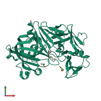
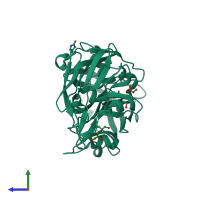
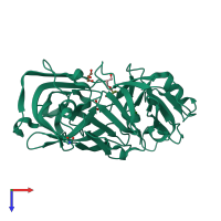
3c9x

X-ray diffraction
1.7Å resolution

Crystal structure of Trichoderma reesei aspartic proteinase

Released:

Function and Biology Details

Reaction catalysed:
Hydrolysis of proteins with broad specificity. Generally favors hydrophobic residues in P1 and P1', but also accepts Lys in P1, which leads to activation of trypsinogen. Does not clot milk.
Biological process:
Cellular component:
  • not assigned

Structure analysis Details

Assembly composition:
monomeric (preferred)
PDBe Complex ID:
PDB-CPX-174124 (preferred)
Entry contents:
1 distinct polypeptide molecule
Macromolecule:
Peptidase A1 domain-containing protein Chain: A
Molecule details ›
Chain: A
Length: 329 amino acids
Theoretical weight: 34.4 KDa
Source organism: Trichoderma reesei
UniProt:
  • Canonical: Q2WBH2 (Residues: 79-407; Coverage: 85%)
Gene name: proA
Sequence domains: Eukaryotic aspartyl protease
Structure domains: Acid Proteases

Ligands and Environments

1 bound ligand:
1 modified residue:

Experiments and Validation Details

Entry percentile scores
X-ray source: LNLS BEAMLINE D03B-MX1
Spacegroup: P43212
Unit cell:
a: 74.171Å b: 74.171Å c: 161.53Å
α: 90° β: 90° γ: 90°
R-values:
R R work R free
0.182 0.179 0.213