3ca7

X-ray diffraction
1.5Å resolution

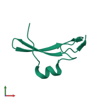
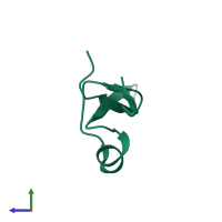
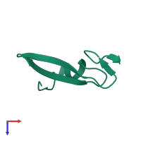
High Resolution Crystal Structure of the EGF domain of Spitz

Released:
Source organism: Drosophila melanogaster
Primary publication:
Structural basis for EGFR ligand sequestration by Argos.
Nature 453 1271-5 (2008)
PMID: 18500331

Function and Biology Details

Biochemical function:
Cellular component:
  • not assigned

Structure analysis Details

Assembly composition:
monomeric (preferred)
Assembly name:
PDBe Complex ID:
PDB-CPX-169515 (preferred)
Entry contents:
1 distinct polypeptide molecule
Macromolecule:
Protein spitz Chain: A
Molecule details ›
Chain: A
Length: 52 amino acids
Theoretical weight: 6.01 KDa
Source organism: Drosophila melanogaster
Expression system: Drosophila melanogaster
UniProt:
  • Canonical: Q01083 (Residues: 76-127; Coverage: 25%)
Gene names: CG10334, spi
Structure domains: Laminin

Ligands and Environments

No bound ligands
No modified residues

Experiments and Validation Details

Entry percentile scores
X-ray source: APS BEAMLINE 23-ID-D
Spacegroup: C2
Unit cell:
a: 58.305Å b: 36.154Å c: 25.362Å
α: 90° β: 103.09° γ: 90°
R-values:
R R work R free
0.201 0.2 0.236
Expression system: Drosophila melanogaster