


Function and Biology Details
Reaction catalysed:
all-trans-violaxanthin + 2 L-ascorbate = all-trans-zeaxanthin +2 L-dehydroascorbate + 2 H2O.
Biochemical function:
Biological process:
Cellular component:
- not assigned
Sequence domains:
Structure analysis Details
Assembly composition:
monomeric (preferred)
Assembly name:
Violaxanthin de-epoxidase, chloroplastic (preferred)
PDBe Complex ID:
PDB-CPX-174441 (preferred)
Entry contents:
1 distinct polypeptide molecule
Macromolecule:
Violaxanthin de-epoxidase, chloroplastic
Molecule details ›
Chains: A, B
Length: 185 amino acids
Theoretical weight: 21.34 KDa
Source organism: Arabidopsis thaliana
Expression system: Escherichia coli
UniProt:
Sequence domains: VDE lipocalin domain
Structure domains: Lipocalin
Length: 185 amino acids
Theoretical weight: 21.34 KDa
Source organism: Arabidopsis thaliana
Expression system: Escherichia coli
UniProt:
- Canonical:
 Q39249 (Residues: 191-366; Coverage: 38%)
Sequence domains: VDE lipocalin domain
Structure domains: Lipocalin
Ligands and Environments
No bound ligands
No modified residues
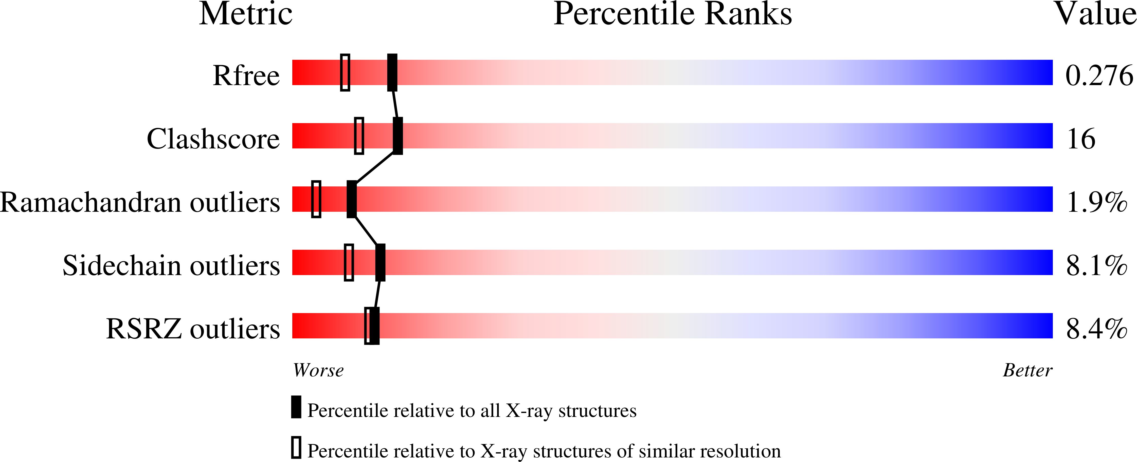
Experiments and Validation Details
wwPDB Validation report is not available for this entry.
X-ray source:
ESRF BEAMLINE ID23-1
Spacegroup:
P1
Expression system: Escherichia coli
