


Function and Biology Details
Biochemical function:
Biological process:
Cellular component:
- not assigned
Sequence domains:
Structure analysis Details
Assembly composition:
homo dimer (preferred)
Assembly name:
Cyanovirin-N (preferred)
PDBe Complex ID:
PDB-CPX-160490 (preferred)
Entry contents:
1 distinct polypeptide molecule
Macromolecule:
Cyanovirin-N
Molecule details ›
Chains: A, B
Length: 101 amino acids
Theoretical weight: 10.69 KDa
Source organism: Nostoc ellipsosporum
Expression system: Escherichia coli
UniProt:
Structure domains: Cyanovirin-N
Length: 101 amino acids
Theoretical weight: 10.69 KDa
Source organism: Nostoc ellipsosporum
Expression system: Escherichia coli
UniProt:
- Canonical:
 P81180 (Residues: 1-101; Coverage: 100%)
Structure domains: Cyanovirin-N
Ligands and Environments
No bound ligands
No modified residues
Experiments and Validation Details
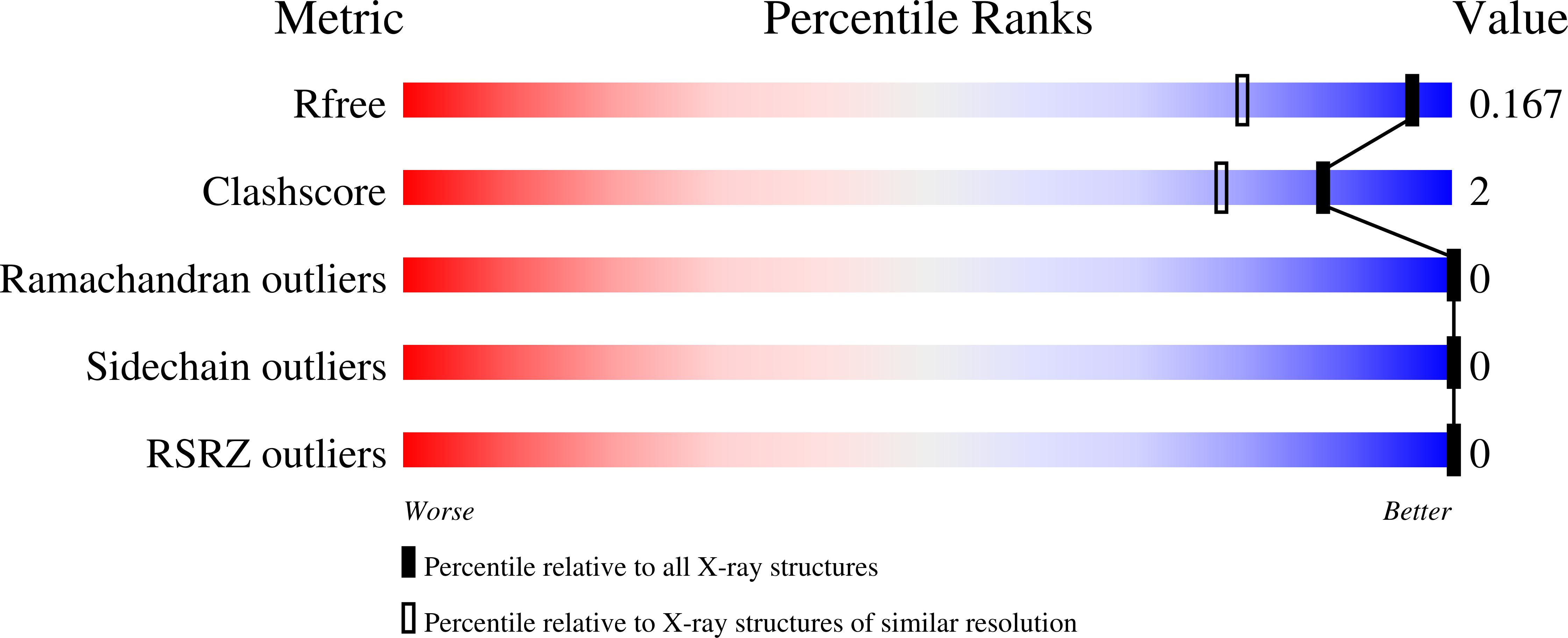
wwPDB Validation report is not available for this entry.
X-ray source:
RIGAKU FR-E SUPERBRIGHT
Spacegroup:
P212121
Expression system: Escherichia coli
