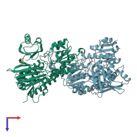
Function and Biology Details
Reactions catalysed:
(R)-2-hydroxyglutarate + NAD(+) = 2-oxoglutarate + NADH
3-phospho-D-glycerate + NAD(+) = 3-phosphonooxypyruvate + NADH
Biochemical function:
Biological process:
Cellular component:
Sequence domains:
- ACT domain
- Allosteric substrate binding domain superfamily
- D-isomer specific 2-hydroxyacid dehydrogenase, NAD-binding domain conserved site
- ACT-like domain
- D-3-phosphoglycerate dehydrogenase, ASB domain
- D-isomer specific 2-hydroxyacid dehydrogenase, NAD-binding domain conserved site 1
- D-isomer specific 2-hydroxyacid dehydrogenase, NAD-binding domain
- NAD(P)-binding domain superfamily
2 more domains
Structure analysis Details
Assembly composition:
homo tetramer (preferred)
Assembly name:
D-3-phosphoglycerate dehydrogenase (preferred)
PDBe Complex ID:
PDB-CPX-161904 (preferred)
Entry contents:
1 distinct polypeptide molecule
Macromolecule:


