3dvt

X-ray diffraction
2.3Å resolution

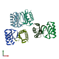
Biochemical and structural characterization of the PAK1- LC8 interaction

Released:
Model geometry
Fit model/data

Function and Biology Details

Structure analysis Details

Assembly composition:
homo dimer (preferred)
PDBe Complex ID:
PDB-CPX-173172 (preferred)
Entry contents:
1 distinct polypeptide molecule
Macromolecule:
Dynein light chain 1, cytoplasmic Chains: A, B, C, D, E, F
Molecule details ›
Chains: A, B, C, D, E, F
Length: 91 amino acids
Theoretical weight: 10.64 KDa
Source organism: Drosophila melanogaster
Expression system: Escherichia coli
UniProt:
  • Canonical: Q24117 (Residues: 1-89; Coverage: 100%)
Gene names: CG6998, Cdlc1, ctp, ddlc1
Sequence domains: Dynein light chain type 1
Structure domains: Protein Inhibitor Of Neuronal Nitric Oxide Synthase;

Ligands and Environments

No bound ligands
No modified residues

Experiments and Validation Details

wwPDB Validation report is not available for this entry.
X-ray source: NSLS BEAMLINE X4C
Spacegroup: P1
Unit cell:
a: 36.482Å b: 44.871Å c: 84.825Å
α: 79.62° β: 77.54° γ: 88.03°
R-values:
R R work R free
0.193 0.189 0.258
Expression system: Escherichia coli