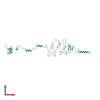
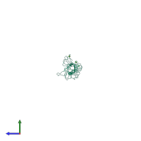
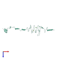
3eka

X-ray diffraction
3.1Å resolution

Crystal structure of the complex of hyaluranidase trimer with ascorbic acid at 3.1 A resolution reveals the locations of three binding sites

Released:

Function and Biology Details

Reaction catalysed:
Cleaves hyaluronate chains at a beta-D-GlcNAc-(1->4)-beta-D-GlcA bond, ultimately breaking the polysaccharide down to 3-(4-deoxy-beta-D-gluc-4-enuronosyl)-N-acetyl-D-glucosamine
Biochemical function:
Cellular component:
  • not assigned

Structure analysis Details

Assembly composition:
homo trimer (preferred)
PDBe Complex ID:
PDB-CPX-189445 (preferred)
Entry contents:
1 distinct polypeptide molecule
Macromolecule:
Major tropism determinant N-terminal domain-containing protein Chain: A
Molecule details ›
Chain: A
Length: 332 amino acids
Theoretical weight: 35.8 KDa
Source organism: Streptococcus pyogenes
Expression system: Escherichia coli
UniProt:
  • Canonical: Q9A0M7 (Residues: 7-337; Coverage: 98%)
Gene names: SPy_0701, hylP1
Sequence domains:

Ligands and Environments

1 bound ligand:
No modified residues

Experiments and Validation Details

Entry percentile scores
X-ray source: EMBL/DESY, HAMBURG BEAMLINE X31
Spacegroup: R32
Unit cell:
a: 58.502Å b: 58.502Å c: 583.54Å
α: 90° β: 90° γ: 120°
R-values:
R R work R free
0.212 0.197 0.234
Expression system: Escherichia coli