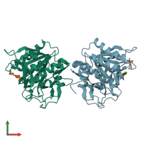
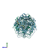
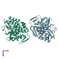
3f7o

X-ray diffraction
2.2Å resolution

Crystal structure of Cuticle-Degrading Protease from Paecilomyces lilacinus (PL646)

Released:

Function and Biology Details

Structure analysis Details

Assembly composition:
hetero dimer (preferred)
PDBe Complex ID:
PDB-CPX-163537 (preferred)
Entry contents:
2 distinct polypeptide molecules
Macromolecules (2 distinct):
Peptidase S8/S53 domain-containing protein Chains: A, B
Molecule details ›
Chains: A, B
Length: 284 amino acids
Theoretical weight: 28.74 KDa
Source organism: Purpureocillium lilacinum
UniProt:
  • Canonical: Q01471 (Residues: 84-367; Coverage: 77%)
Sequence domains: Subtilase family
Structure domains: Peptidase S8/S53 domain
(MSU)(ALA)(ALA)(PRO)(VAL) Chains: X, Y
Molecule details ›
Chains: X, Y
Length: 5 amino acids
Theoretical weight: 471 Da
Source organism: synthetic construct
Expression system: Not provided

Ligands and Environments

1 bound ligand:
No modified residues

Experiments and Validation Details

Entry percentile scores
X-ray source: RIGAKU MICROMAX-007
Spacegroup: P21
Unit cell:
a: 65.108Å b: 62.536Å c: 67.562Å
α: 90° β: 92.76° γ: 90°
R-values:
R R work R free
0.231 0.229 0.258
Expression system: Not provided