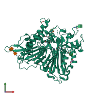
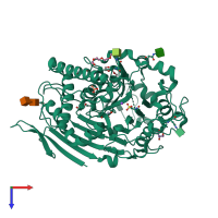
3fbx

X-ray diffraction
2.4Å resolution

Crystal structure of the lysosomal 66.3 kDa protein from mouse solved by S-SAD

Released:
Source organism: Mus musculus
Primary publication:
De novo sulfur SAD phasing of the lysosomal 66.3 kDa protein from mouse.
Acta Crystallogr D Biol Crystallogr 65 220-8 (2009)
PMID: 19237744

Function and Biology Details

Structure analysis Details

Assembly composition:
homo dimer (preferred)
PDBe Complex ID:
PDB-CPX-174812 (preferred)
Entry contents:
1 distinct polypeptide molecule
Macromolecules (2 distinct):
Putative phospholipase B-like 2 Chain: A
Molecule details ›
Chain: A
Length: 559 amino acids
Theoretical weight: 63.23 KDa
Source organism: Mus musculus
Expression system: Homo sapiens
UniProt:
  • Canonical: Q3TCN2 (Residues: 47-594; Coverage: 100%)
Gene name: Plbd2
Sequence domains: Phospholipase B
Structure domains: Penicillin V Acylase; Chain A

Ligands and Environments

Carbohydrate polymer : NEW Components: NAG
1 modified residue:

Experiments and Validation Details

Entry percentile scores
X-ray source: BESSY BEAMLINE 14.2
Spacegroup: C2
Unit cell:
a: 148.804Å b: 89.672Å c: 64.954Å
α: 90° β: 98.67° γ: 90°
R-values:
R R work R free
0.158 0.156 0.198
Expression system: Homo sapiens