


Function and Biology Details
Biochemical function:
Biological process:
- not assigned
Cellular component:
Sequence domains:
Structure analysis Details
Assembly composition:
homo dimer (preferred)
Assembly name:
Nudix hydrolase domain-containing protein (preferred)
PDBe Complex ID:
PDB-CPX-191777 (preferred)
Entry contents:
1 distinct polypeptide molecule
Macromolecule:
Nudix hydrolase domain-containing protein
Molecule details ›
Chains: A, B
Length: 188 amino acids
Theoretical weight: 21.8 KDa
Source organism: Alkalihalobacillus halodurans
Expression system: Escherichia coli
UniProt:
Sequence domains: NUDIX domain
Structure domains: Nucleoside Triphosphate Pyrophosphohydrolase
Length: 188 amino acids
Theoretical weight: 21.8 KDa
Source organism: Alkalihalobacillus halodurans
Expression system: Escherichia coli
UniProt:
- Canonical:
 Q9K704 (Residues: 2-157; Coverage: 98%)
Sequence domains: NUDIX domain
Structure domains: Nucleoside Triphosphate Pyrophosphohydrolase
Ligands and Environments
No bound ligands
No modified residues
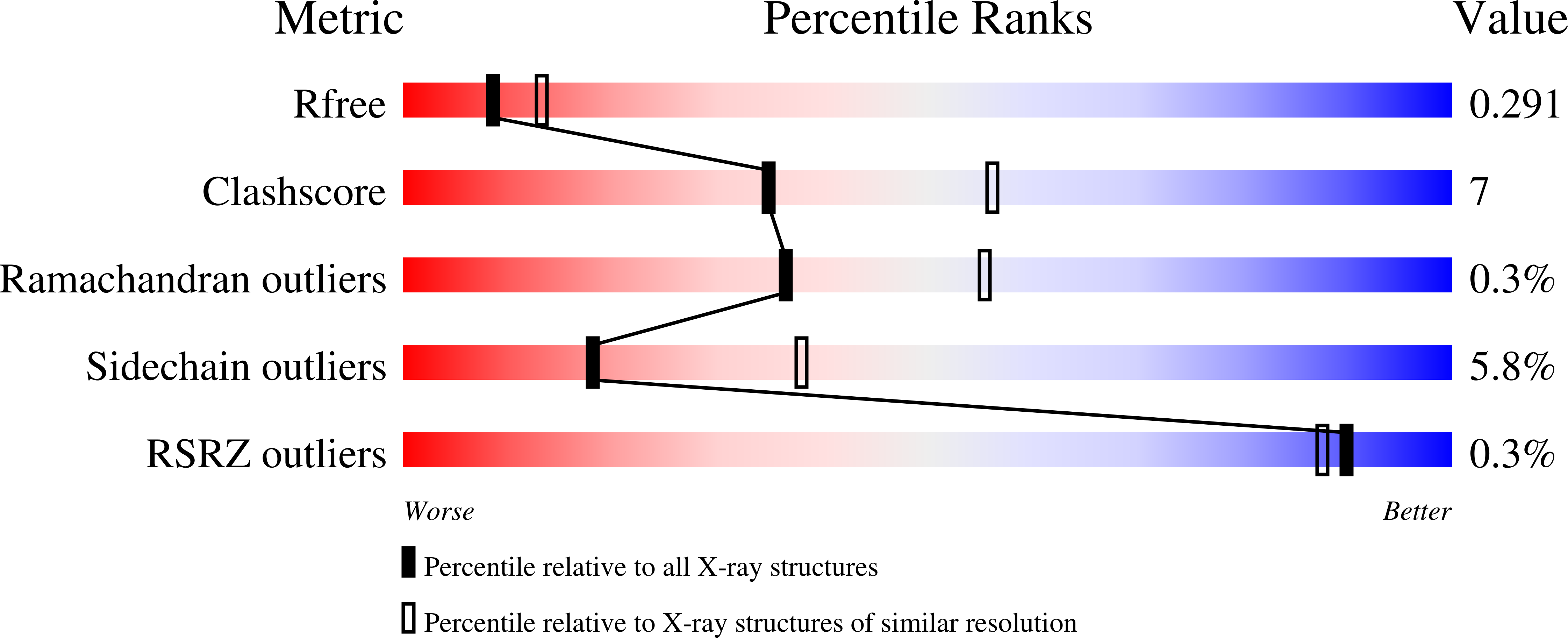
Experiments and Validation Details
wwPDB Validation report is not available for this entry.
X-ray source:
APS BEAMLINE 31-ID
Spacegroup:
P212121
Expression system: Escherichia coli
