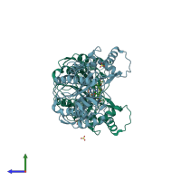
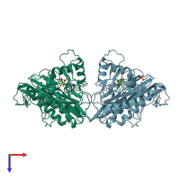
3fmq

X-ray diffraction
2.5Å resolution

Crystal structure of an Encephalitozoon cuniculi methionine aminopeptidase type 2 with angiogenesis inhibitor fumagillin bound

Released:

Function and Biology Details

Structure analysis Details

Assembly composition:
monomeric (preferred)
PDBe Complex ID:
PDB-CPX-186085 (preferred)
Entry contents:
1 distinct polypeptide molecule
Macromolecule:
Methionine aminopeptidase 2 Chains: A, B
Molecule details ›
Chains: A, B
Length: 358 amino acids
Theoretical weight: 40.03 KDa
Source organism: Encephalitozoon cuniculi
Expression system: Spodoptera frugiperda
UniProt:
  • Canonical: Q8SR45 (Residues: 1-358; Coverage: 100%)
Gene names: ECU10_0750, MAP2
Sequence domains: Metallopeptidase family M24
Structure domains:

Ligands and Environments

3 bound ligands:
No modified residues

Experiments and Validation Details

Entry percentile scores
X-ray source: NSLS BEAMLINE X29A
Spacegroup: P21
Unit cell:
a: 62.008Å b: 94.238Å c: 66.325Å
α: 90° β: 99.43° γ: 90°
R-values:
R R work R free
0.176 0.173 0.229
Expression system: Spodoptera frugiperda