3g5i

X-ray diffraction
2.1Å resolution

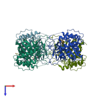
Crystal Structure of the E.coli RihA pyrimidine nucleosidase bound to a iminoribitol-based inhibitor

Released:

Function and Biology Details

Structure analysis Details

Assembly composition:
homo tetramer (preferred)
PDBe Complex ID:
PDB-CPX-154394 (preferred)
Entry contents:
1 distinct polypeptide molecule
Macromolecule:
Pyrimidine-specific ribonucleoside hydrolase RihA Chains: A, B, C, D
Molecule details ›
Chains: A, B, C, D
Length: 312 amino acids
Theoretical weight: 33.87 KDa
Source organism: Escherichia coli K-12
Expression system: Escherichia coli
UniProt:
  • Canonical: P41409 (Residues: 2-311; Coverage: 100%)
Gene names: JW0646, b0651, rihA, ybeK
Sequence domains: Inosine-uridine preferring nucleoside hydrolase
Structure domains: Ribonucleoside hydrolase-like

Ligands and Environments

3 bound ligands:
No modified residues

Experiments and Validation Details

Entry percentile scores
X-ray source: ESRF BEAMLINE ID14-2
Spacegroup: P21
Unit cell:
a: 84.35Å b: 82.91Å c: 93.87Å
α: 90° β: 112.15° γ: 90°
R-values:
R R work R free
0.205 0.203 0.241
Expression system: Escherichia coli