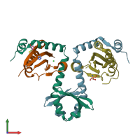
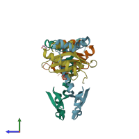
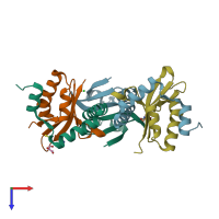
3g5o

X-ray diffraction
2Å resolution

The crystal structure of the toxin-antitoxin complex RelBE2 (Rv2865-2866) from Mycobacterium tuberculosis

Released:

Function and Biology Details

Structure analysis Details

Assembly composition:
hetero tetramer (preferred)
PDBe Complex ID:
PDB-CPX-128484 (preferred)
Entry contents:
2 distinct polypeptide molecules
Macromolecules (2 distinct):
Antitoxin RelF Chains: A, D
Molecule details ›
Chains: A, D
Length: 108 amino acids
Theoretical weight: 11.86 KDa
Source organism: Mycobacterium tuberculosis H37Rv
Expression system: Escherichia coli
UniProt:
  • Canonical: O33347 (Residues: 1-93; Coverage: 100%)
Gene names: Rv2865, relB2, relF
Sequence domains: Antitoxin Phd_YefM, type II toxin-antitoxin system
Structure domains:
Toxin RelG Chains: B, C
Molecule details ›
Chains: B, C
Length: 102 amino acids
Theoretical weight: 12.08 KDa
Source organism: Mycobacterium tuberculosis H37Rv
Expression system: Escherichia coli
UniProt:
  • Canonical: O33348 (Residues: 2-87; Coverage: 99%)
Gene names: Rv2866, relE2, relG
Sequence domains: ParE toxin of type II toxin-antitoxin system, parDE
Structure domains: RelE-like

Ligands and Environments

No modified residues

Experiments and Validation Details

Entry percentile scores
X-ray source: APS BEAMLINE 24-ID-C, ALS BEAMLINE 8.2.1
Spacegroup: P212121
Unit cell:
a: 75.54Å b: 79.481Å c: 86.278Å
α: 90° β: 90° γ: 90°
R-values:
R R work R free
0.205 0.202 0.246
Expression system: Escherichia coli