


Function and Biology Details
Biochemical function:
Biological process:
- not assigned
Cellular component:
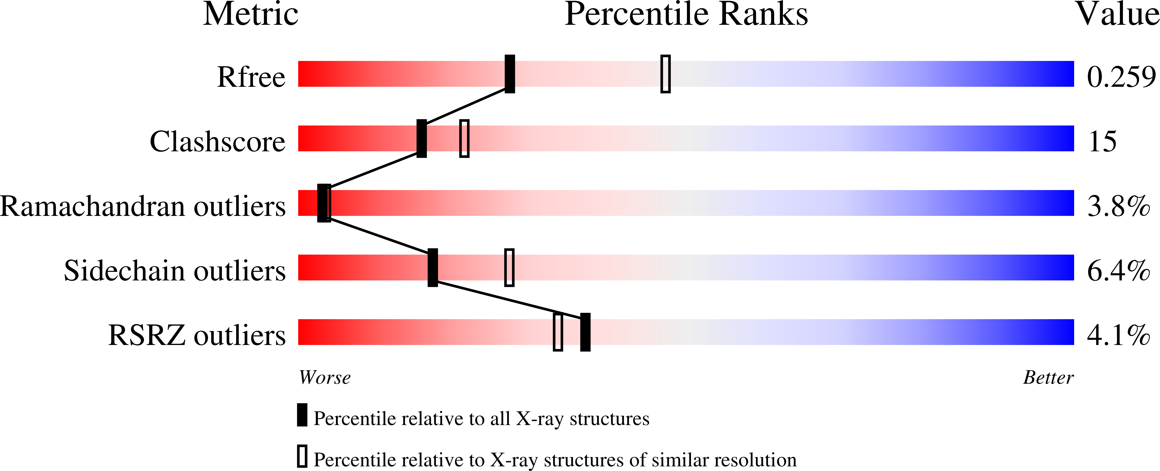
Structure analysis Details
Assemblies composition:
Assembly name:
Chaperone protein IpgC (preferred)
PDBe Complex ID:
PDB-CPX-141073 (preferred)
Entry contents:
2 distinct polypeptide molecules
Macromolecules (2 distinct):
Chaperone protein IpgC
Molecule details ›
Chains: A, B
Length: 151 amino acids
Theoretical weight: 17.21 KDa
Source organism: Shigella flexneri
Expression system: Escherichia coli
UniProt:
Sequence domains: Tetratricopeptide repeat
Structure domains: Tetratricopeptide repeat domain
Length: 151 amino acids
Theoretical weight: 17.21 KDa
Source organism: Shigella flexneri
Expression system: Escherichia coli
UniProt:
- Canonical:
 P0A2U4 (Residues: 1-151; Coverage: 97%)
Sequence domains: Tetratricopeptide repeat
Structure domains: Tetratricopeptide repeat domain
Type 3 secretion system translocon protein SctE
Molecule details ›
Chain: P
Length: 78 amino acids
Theoretical weight: 8.3 KDa
Source organism: Shigella flexneri
Expression system: Escherichia coli
UniProt:
Length: 78 amino acids
Theoretical weight: 8.3 KDa
Source organism: Shigella flexneri
Expression system: Escherichia coli
UniProt:
- Canonical: P18011 (Residues: 16-72; Coverage: 10%)


