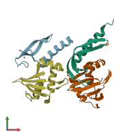
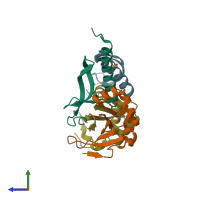
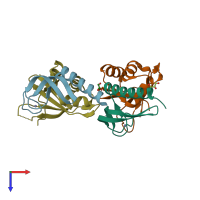
3hi2

X-ray diffraction
2Å resolution

Structure of the N-terminal domain of the E. coli antitoxin MqsA (YgiT/b3021) in complex with the E. coli toxin MqsR (YgiU/b3022)

Released:

Function and Biology Details

Structure analysis Details

Assembly composition:
hetero dimer (preferred)
PDBe Complex ID:
PDB-CPX-175149 (preferred)
Entry contents:
2 distinct polypeptide molecules
Macromolecules (2 distinct):
Antitoxin MqsA Chains: A, C
Molecule details ›
Chains: A, C
Length: 76 amino acids
Theoretical weight: 8.52 KDa
Source organism: Escherichia coli K-12
Expression system: Escherichia coli
UniProt:
  • Canonical: Q46864 (Residues: 1-76; Coverage: 58%)
Gene names: JW2989, b3021, mqsA, ygiT
Sequence domains: Antitoxin component of bacterial toxin-antitoxin system, MqsA
Structure domains: Ubiquitin-like (UB roll)
mRNA interferase toxin MqsR Chains: B, D
Molecule details ›
Chains: B, D
Length: 101 amino acids
Theoretical weight: 11.53 KDa
Source organism: Escherichia coli K-12
Expression system: Escherichia coli
UniProt:
  • Canonical: Q46865 (Residues: 1-98; Coverage: 100%)
Gene names: JW2990, b3022, mqsR, ygiU
Sequence domains: Motility quorum-sensing regulator, toxin of MqsA
Structure domains: YaeB-like fold

Ligands and Environments

2 bound ligands:
No modified residues

Experiments and Validation Details

Entry percentile scores
X-ray source: NSLS BEAMLINE X6A
Spacegroup: P41212
Unit cell:
a: 63.234Å b: 63.234Å c: 194.886Å
α: 90° β: 90° γ: 90°
R-values:
R R work R free
0.207 0.205 0.248
Expression system: Escherichia coli