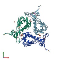
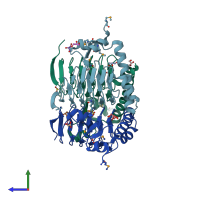
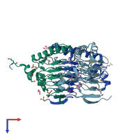
3hjj

X-ray diffraction
2.15Å resolution

Crystal Structure of Maltose O-acetyltransferase from Bacillus anthracis

Released:
Source organism: Bacillus anthracis
Entry authors: Kim Y, Maltseva N, Papazisi L, Anderson W, Joachimiak A, Center for Structural Genomics of Infectious Diseases (CSGID)

Function and Biology Details

Reaction catalysed:
Acetyl-CoA + maltose = CoA + acetyl-maltose
Biochemical function:
Biological process:
  • not assigned
Cellular component:

Structure analysis Details

Assembly composition:
homo trimer (preferred)
PDBe Complex ID:
PDB-CPX-105680 (preferred)
Entry contents:
1 distinct polypeptide molecule
Macromolecule:
Maltose/galactoside acetyltransferase domain-containing protein Chains: A, B, C
Molecule details ›
Chains: A, B, C
Length: 190 amino acids
Theoretical weight: 20.82 KDa
Source organism: Bacillus anthracis
Expression system: Escherichia coli
UniProt:
  • Canonical: A0A6L7H2S4 (Residues: 1-187; Coverage: 100%)
Gene names: GBAA_3402, maa
Sequence domains:
Structure domains: Hexapeptide repeat proteins

Ligands and Environments

3 bound ligands:
1 modified residue:

Experiments and Validation Details

Entry percentile scores
X-ray source: APS BEAMLINE 19-BM
Spacegroup: P41212
Unit cell:
a: 123.01Å b: 123.01Å c: 144.003Å
α: 90° β: 90° γ: 90°
R-values:
R R work R free
0.166 0.165 0.194
Expression system: Escherichia coli