


Function and Biology Details
Biochemical function:
- not assigned
Biological process:
- not assigned
Cellular component:
- not assigned
Sequence domain:
Structure analysis Details
Assembly composition:
homo dimer (preferred)
Assembly name:
HTH lysR-type domain-containing protein (preferred)
PDBe Complex ID:
PDB-CPX-181780 (preferred)
Entry contents:
1 distinct polypeptide molecule
Macromolecule:
HTH lysR-type domain-containing protein
Molecule details ›
Chains: A, B
Length: 232 amino acids
Theoretical weight: 26.69 KDa
Source organism: Porphyromonas gingivalis
Expression system: Escherichia coli
UniProt:
Sequence domains: LysR substrate binding domain
Structure domains: Periplasmic binding protein-like II
Length: 232 amino acids
Theoretical weight: 26.69 KDa
Source organism: Porphyromonas gingivalis
Expression system: Escherichia coli
UniProt:
- Canonical:
 Q7MXD3 (Residues: 81-308; Coverage: 74%)
Sequence domains: LysR substrate binding domain
Structure domains: Periplasmic binding protein-like II
Ligands and Environments
No bound ligands
No modified residues
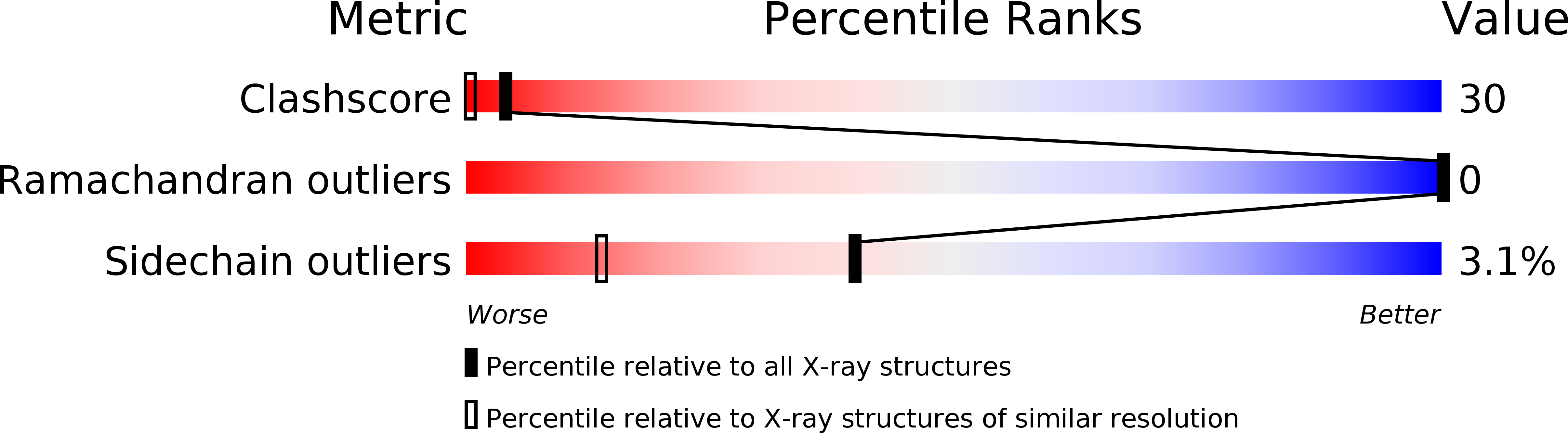
Experiments and Validation Details

X-ray source:
RIGAKU MICROMAX-007
Spacegroup:
P1
Expression system: Escherichia coli