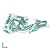
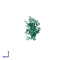
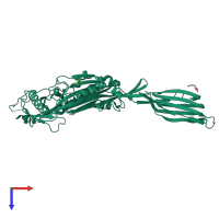
3hvn

X-ray diffraction
2.85Å resolution

Crystal structure of cytotoxin protein suilysin from Streptococcus suis

Released:
Source organism: Streptococcus suis 05ZYH33
Primary publication:
Crystal structure of cytotoxin protein suilysin from Streptococcus suis.
Protein Cell 1 96-105 (2010)
PMID: 21204001

Function and Biology Details

Structure analysis Details

Assembly composition:
monomeric (preferred)
PDBe Complex ID:
PDB-CPX-237461 (preferred)
Entry contents:
1 distinct polypeptide molecule
Macromolecule:
Thiol-activated cytolysin Chain: A
Molecule details ›
Chain: A
Length: 508 amino acids
Theoretical weight: 56.1 KDa
Source organism: Streptococcus suis 05ZYH33
Expression system: Escherichia coli BL21(DE3)
UniProt:
  • Canonical: Q55996 (Residues: 2-497; Coverage: 100%)
Gene names: ERS132356_00692, ERS132441_00352, ERS132462_00946, Q7V66_08590, ply, sly
Sequence domains:
Structure domains:

Ligands and Environments

2 bound ligands:
No modified residues

Experiments and Validation Details

Entry percentile scores
X-ray source: PHOTON FACTORY BEAMLINE BL-5A, PHOTON FACTORY BEAMLINE BL-17A
Spacegroup: P3121
Unit cell:
a: 95.72Å b: 95.72Å c: 117.046Å
α: 90° β: 90° γ: 120°
R-values:
R R work R free
0.28 0.279 0.294
Expression system: Escherichia coli BL21(DE3)