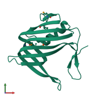
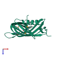
3hwu

X-ray diffraction
1.3Å resolution

Crystal structure of Putative DNA-binding protein (YP_299413.1) from Ralstonia eutrophA JMP134 at 1.30 A resolution

Released:
Entry author: Joint Center for Structural Genomics (JCSG)

Function and Biology Details

Biochemical function:
  • not assigned
Biological process:
  • not assigned
Cellular component:
  • not assigned

Structure analysis Details

Assembly composition:
homo trimer (preferred)
PDBe Complex ID:
PDB-CPX-175188 (preferred)
Entry contents:
1 distinct polypeptide molecule
Macromolecule:
PPC domain-containing protein Chain: A
Molecule details ›
Chain: A
Length: 147 amino acids
Theoretical weight: 16.39 KDa
Source organism: Cupriavidus pinatubonensis JMP134
Expression system: Escherichia coli
UniProt:
  • Canonical: Q46QL5 (Residues: 28-173; Coverage: 100%)
Gene name: Reut_B5223
Sequence domains: Plants and Prokaryotes Conserved (PCC) domain
Structure domains: Hypothetical protein, similar to alpha- acetolactate decarboxylase; domain 2

Ligands and Environments

No bound ligands
1 modified residue:

Experiments and Validation Details

Entry percentile scores
X-ray source: SSRL BEAMLINE BL9-2
Spacegroup: R3
Unit cell:
a: 53.897Å b: 53.897Å c: 111.1Å
α: 90° β: 90° γ: 120°
R-values:
R R work R free
0.121 0.119 0.14
Expression system: Escherichia coli