3ifs

X-ray diffraction
2Å resolution

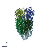
2.0 Angstrom Resolution Crystal Structure of Glucose-6-phosphate Isomerase (pgi) from Bacillus anthracis.

Released:
Entry authors: Minasov G, Wawrzak Z, Onopriyenko O, Gordon E, Peterson SN, Savchenko A, Anderson WF, Center for Structural Genomics of Infectious Diseases (CSGID)

Function and Biology Details

Reaction catalysed:
Alpha-D-glucose 6-phosphate = beta-D-fructofuranose 6-phosphate
Biochemical function:
Cellular component:

Structure analysis Details

Assemblies composition:
homo dimer
homo tetramer (preferred)
PDBe Complex ID:
PDB-CPX-182562 (preferred)
Entry contents:
1 distinct polypeptide molecule
Macromolecule:
Glucose-6-phosphate isomerase Chains: A, B, C, D, E, F
Molecule details ›
Chains: A, B, C, D, E, F
Length: 453 amino acids
Theoretical weight: 51.1 KDa
Source organism: Bacillus anthracis str. 'Ames Ancestor'
Expression system: Escherichia coli
UniProt:
  • Canonical: Q81K75 (Residues: 1-450; Coverage: 100%)
Gene names: BAS4767, BA_5130, GBAA_5130, pgi
Sequence domains: Phosphoglucose isomerase
Structure domains: Glucose-6-phosphate isomerase like protein; domain 1

Ligands and Environments

1 modified residue:

Experiments and Validation Details

Entry percentile scores
X-ray source: APS BEAMLINE 21-ID-G
Spacegroup: P21212
Unit cell:
a: 111.802Å b: 303.64Å c: 72.087Å
α: 90° β: 90° γ: 90°
R-values:
R R work R free
0.143 0.14 0.19
Expression system: Escherichia coli