3irc

X-ray diffraction
2.25Å resolution

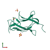
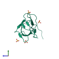
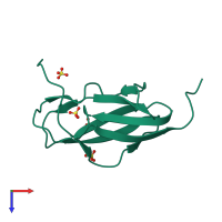
Crystal structure analysis of dengue-1 envelope protein domain III

Released:
Source organism: Dengue virus 1
Entry authors: Nelson CA, Kim T, Warren JT, Chruszcz M, Minor W, Fremont DH, Center for Structural Genomics of Infectious Diseases (CSGID)

Function and Biology Details

Reactions catalysed:
Selective hydrolysis of -Xaa-Xaa-|-Yaa- bonds in which each of the Xaa can be either Arg or Lys and Yaa can be either Ser or Ala.
NTP + H(2)O = NDP + phosphate
ATP + H(2)O = ADP + phosphate
Biochemical function:
  • not assigned
Biological process:
  • not assigned
Cellular component:
  • not assigned

Structure analysis Details

Assemblies composition:
monomeric (preferred)
homo trimer
Assembly name:
PDBe Complex ID:
PDB-CPX-191578 (preferred)
Entry contents:
1 distinct polypeptide molecule
Macromolecule:
ENVELOPE PROTEIN Chain: A
Molecule details ›
Chain: A
Length: 108 amino acids
Theoretical weight: 11.7 KDa
Source organism: Dengue virus 1
Expression system: Escherichia coli
UniProt:
  • Canonical: Q9J7C6 (Residues: 576-679; Coverage: 3%)
Gene name: POLY
Sequence domains: Flavivirus glycoprotein, immunoglobulin-like domain
Structure domains: Immunoglobulin-like

Ligands and Environments

1 bound ligand:
No modified residues

Experiments and Validation Details

Entry percentile scores
X-ray source: ALS BEAMLINE 4.2.2
Spacegroup: P213
Unit cell:
a: 88.087Å b: 88.087Å c: 88.087Å
α: 90° β: 90° γ: 90°
R-values:
R R work R free
0.212 0.211 0.221
Expression system: Escherichia coli