3iti

X-ray diffraction
1.55Å resolution

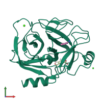
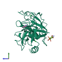
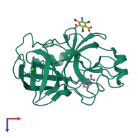
Structure of bovine trypsin with the MAD triangle B3C

Released:
Source organism: Bos taurus
Primary publication:
How to get the magic triangle and the MAD triangle into your protein crystal.
Acta Crystallogr Sect F Struct Biol Cryst Commun 65 1068-70 (2009)
PMID: 19851024

Function and Biology Details

Structure analysis Details

Assembly composition:
monomeric (preferred)
Assembly name:
PDBe Complex ID:
PDB-CPX-133319 (preferred)
Entry contents:
1 distinct polypeptide molecule
Macromolecule:
Serine protease 1 Chain: A
Molecule details ›
Chain: A
Length: 223 amino acids
Theoretical weight: 23.32 KDa
Source organism: Bos taurus
UniProt:
  • Canonical: P00760 (Residues: 24-246; Coverage: 97%)
Gene names: PRSS1, TRP1, TRY1, TRYP1
Sequence domains: Trypsin
Structure domains: Trypsin-like serine proteases

Ligands and Environments

No modified residues

Experiments and Validation Details

Entry percentile scores
X-ray source: MACSCIENCE
Spacegroup: P212121
Unit cell:
a: 53.658Å b: 56.883Å c: 66.808Å
α: 90° β: 90° γ: 90°
R-values:
R R work R free
0.168 not available 0.213