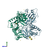
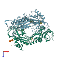
3k1l

X-ray diffraction
3.2Å resolution

Crystal Structure of FANCL

Released:
Source organism: Drosophila melanogaster
Primary publication:
The structure of the catalytic subunit FANCL of the Fanconi anemia core complex.
Nat Struct Mol Biol 17 294-8 (2010)
PMID: 20154706

Function and Biology Details

Reaction catalysed:
S-ubiquitinyl-[E2 ubiquitin-conjugating enzyme]-L-cysteine + [acceptor protein]-L-lysine = [E2 ubiquitin-conjugating enzyme]-L-cysteine + N(6)-ubiquitinyl-[acceptor protein]-L-lysine
Biochemical function:
Biological process:
Cellular component:

Structure analysis Details

Assemblies composition:
monomeric (preferred)
homo hexamer
homo dimer
PDBe Complex ID:
PDB-CPX-186147 (preferred)
Entry contents:
1 distinct polypeptide molecule
Macromolecules (2 distinct):
E3 ubiquitin-protein ligase Fancl Chains: A, B
Molecule details ›
Chains: A, B
Length: 381 amino acids
Theoretical weight: 43.78 KDa
Source organism: Drosophila melanogaster
Expression system: Escherichia coli
UniProt:
  • Canonical: Q8T913 (Residues: 1-381; Coverage: 100%)
Gene names: CG12812, FAL, Fancl
Sequence domains:
Structure domains:

Ligands and Environments

Carbohydrate polymer : NEW Components: GLC
3 bound ligands:
No modified residues

Experiments and Validation Details

Entry percentile scores
X-ray source: DIAMOND BEAMLINE I04
Spacegroup: R32
Unit cell:
a: 188.68Å b: 188.68Å c: 259.36Å
α: 90° β: 90° γ: 120°
R-values:
R R work R free
0.207 0.205 0.247
Expression system: Escherichia coli