


Function and Biology Details
Biochemical function:
Biological process:
Cellular component:
- not assigned
Structure analysis Details
Assembly composition:
homo dimer (preferred)
Assembly name:
Serine protease Rv3671c (preferred)
PDBe Complex ID:
PDB-CPX-161582 (preferred)
Entry contents:
1 distinct polypeptide molecule
Macromolecule:
Serine protease Rv3671c
Molecule details ›
Chain: A
Length: 237 amino acids
Theoretical weight: 24.14 KDa
Source organism: Mycobacterium tuberculosis
Expression system: Escherichia coli
UniProt:
Sequence domains: Trypsin-like peptidase domain
Structure domains: Trypsin-like serine proteases
Length: 237 amino acids
Theoretical weight: 24.14 KDa
Source organism: Mycobacterium tuberculosis
Expression system: Escherichia coli
UniProt:
- Canonical:
 P9WHR9 (Residues: 161-397; Coverage: 60%)
Sequence domains: Trypsin-like peptidase domain
Structure domains: Trypsin-like serine proteases
Ligands and Environments
No bound ligands
No modified residues
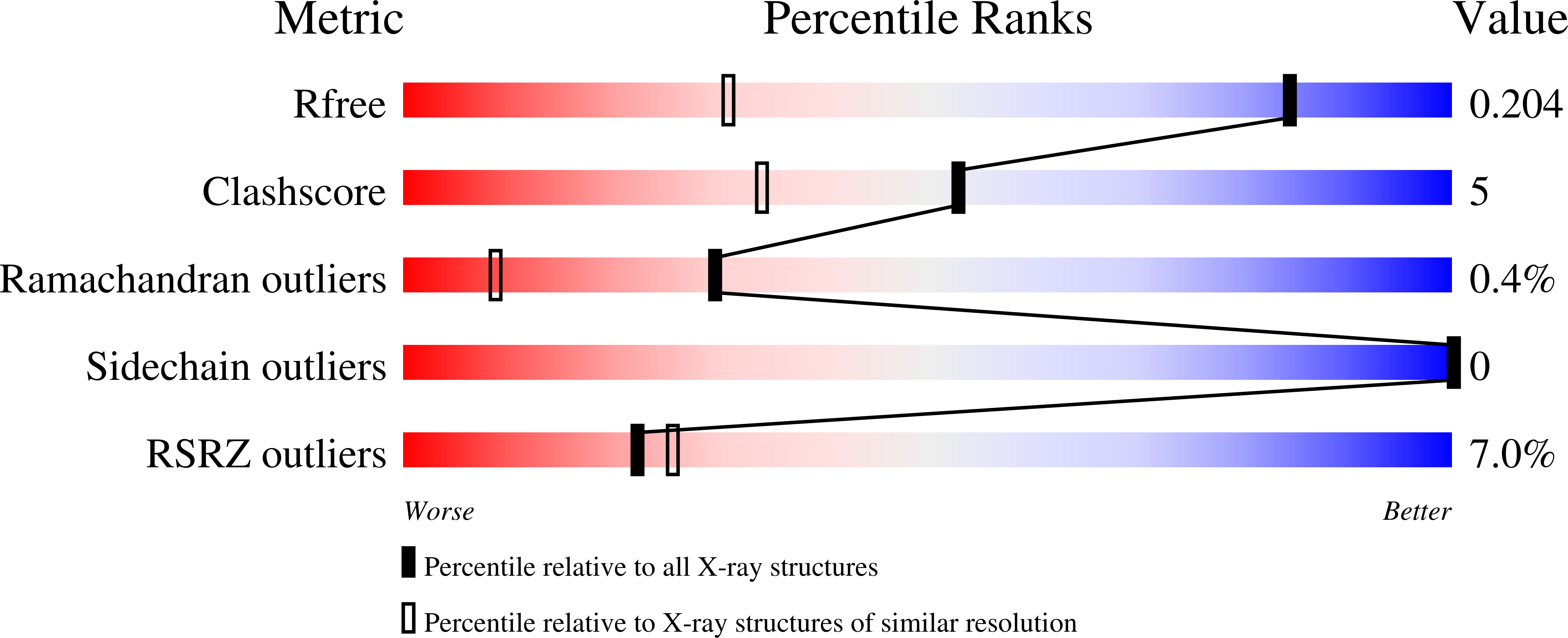
Experiments and Validation Details
wwPDB Validation report is not available for this entry.
X-ray source:
APS BEAMLINE 21-ID-D
Spacegroup:
P3121
Expression system: Escherichia coli
