


Function and Biology Details
Biochemical function:
Biological process:
- not assigned
Cellular component:
- not assigned
Structure analysis Details
Assemblies composition:
Assembly name:
Beta-lactoglobulin, Beta-lactoglobulin-1 (preferred)
PDBe Complex ID:
PDB-CPX-136151 (preferred)
Entry contents:
1 distinct polypeptide molecule
Macromolecule:
Beta-lactoglobulin; Beta-lactoglobulin-1
Molecule details ›
Chains: A, B
Length: 162 amino acids
Theoretical weight: 18.31 KDa
Source organisms: Expression system: Escherichia coli
UniProt:
Sequence domains: Lipocalin / cytosolic fatty-acid binding protein family
Structure domains: Lipocalin
Length: 162 amino acids
Theoretical weight: 18.31 KDa
Source organisms: Expression system: Escherichia coli
UniProt:
- Canonical:
 P02758 (Residues: 19-35, 52-60, 82-85, 95-102, 106-107, 119-121, 128-135, 144-147, 159-162, 173-180; Coverage: 41%)
 - Canonical: P02754 (Residues: 34-49, 59-79, 84-92, 101-103, 106-116, 120-125, 134-141, 146-156, 161-170; Coverage: 59%)
Sequence domains: Lipocalin / cytosolic fatty-acid binding protein family
Structure domains: Lipocalin
Ligands and Environments
No bound ligands
No modified residues
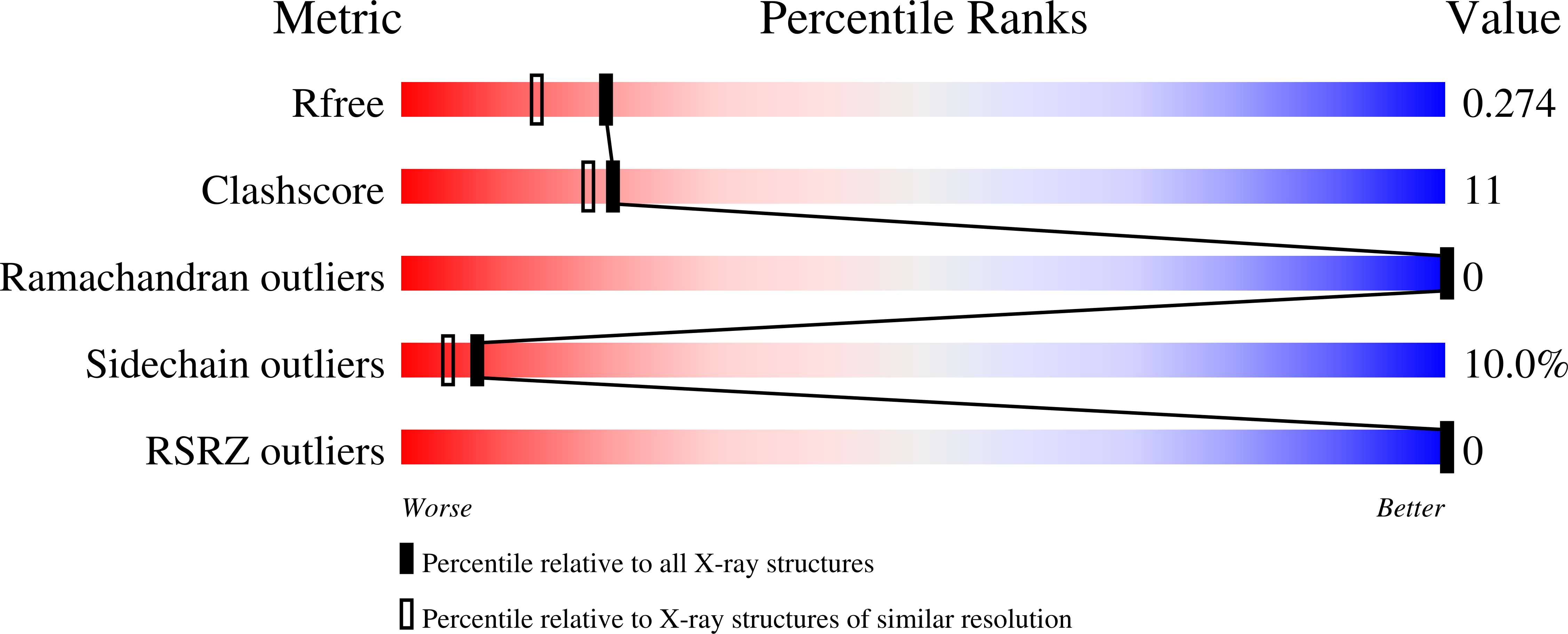
Experiments and Validation Details

X-ray source:
PHOTON FACTORY BEAMLINE AR-NW12A
Spacegroup:
P65
Expression system: Escherichia coli