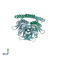
3m65

X-ray diffraction
2.6Å resolution

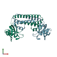
Crystal structure of Bacillus subtilis Lon N-terminal domain

Released:
Primary publication:
Crystal structures of Bacillus subtilis Lon protease.
J Mol Biol 401 653-70 (2010)
PMID: 20600124

Function and Biology Details

Reaction catalysed:
Hydrolysis of proteins in presence of ATP.
Biochemical function:
Biological process:
Cellular component:
  • not assigned

Structure analysis Details

Assemblies composition:
monomeric (preferred)
homo dimer
Assembly name:
PDBe Complex ID:
PDB-CPX-153516 (preferred)
Entry contents:
1 distinct polypeptide molecule
Macromolecule:
Lon protease 1 Chains: A, B
Molecule details ›
Chains: A, B
Length: 209 amino acids
Theoretical weight: 24.01 KDa
Source organism: Bacillus subtilis subsp. subtilis str. 168
Expression system: Escherichia coli
UniProt:
  • Canonical: P37945 (Residues: 1-209; Coverage: 27%)
Gene names: BSU28200, lon1, lonA
Sequence domains: ATP-dependent protease La (LON) substrate-binding domain
Structure domains:

Ligands and Environments

No bound ligands
No modified residues

Experiments and Validation Details

Entry percentile scores
X-ray source: DIAMOND BEAMLINE I02
Spacegroup: P212121
Unit cell:
a: 43.877Å b: 81.759Å c: 115.133Å
α: 90° β: 90° γ: 90°
R-values:
R R work R free
0.195 0.191 0.269
Expression system: Escherichia coli