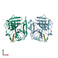
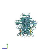
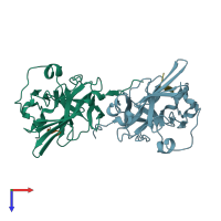
3mmg

X-ray diffraction
1.7Å resolution

Crystal structure of tobacco vein mottling virus protease

Released:

Function and Biology Details

Reactions catalysed:
Nucleoside triphosphate + RNA(n) = diphosphate + RNA(n+1)
Hydrolyzes glutaminyl bonds, and activity is further restricted by preferences for the amino acids in P6 - P1' that vary with the species of potyvirus, e.g. Glu-Xaa-Xaa-Tyr-Xaa-Gln-|-(Ser or Gly) for the enzyme from tobacco etch virus. The natural substrate is the viral polyprotein, but other proteins and oligopeptides containing the appropriate consensus sequence are also cleaved.
Hydrolyzes a Gly-|-Gly bond at its own C-terminus, commonly in the sequence -Tyr-Xaa-Val-Gly-|-Gly, in the processing of the potyviral polyprotein.
Biochemical function:
Biological process:
Cellular component:
  • not assigned

Structure analysis Details

Assemblies composition:
hetero dimer (preferred)
hetero tetramer
Assembly name:
PDBe Complex ID:
PDB-CPX-140760 (preferred)
Entry contents:
2 distinct polypeptide molecules
Macromolecules (2 distinct):
Nuclear inclusion protein A Chains: A, B
Molecule details ›
Chains: A, B
Length: 241 amino acids
Theoretical weight: 27.06 KDa
Source organism: Tobacco vein mottling virus
Expression system: Escherichia coli BL21(DE3)
UniProt:
  • Canonical: P09814 (Residues: 2002-2242; Coverage: 8%)
Sequence domains: Peptidase family C4
Structure domains: Trypsin-like serine proteases
Capsid protein Chains: C, D
Molecule details ›
Chains: C, D
Length: 8 amino acids
Theoretical weight: 982 Da
Source organism: Tobacco vein mottling virus
Expression system: Not provided
UniProt:
  • Canonical: P09814 (Residues: 2753-2760; Coverage: 0%)

Ligands and Environments

1 bound ligand:
No modified residues

Experiments and Validation Details

Entry percentile scores
X-ray source: APS BEAMLINE 22-ID
Spacegroup: P212121
Unit cell:
a: 76.476Å b: 77.563Å c: 78.463Å
α: 90° β: 90° γ: 90°
R-values:
R R work R free
0.176 0.175 0.21
Expression systems:
  • Escherichia coli BL21(DE3)
  • Not provided