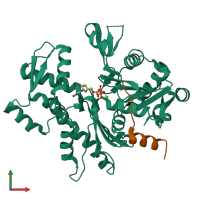
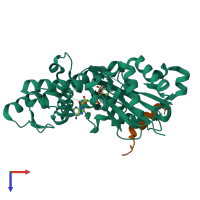
3mn7

X-ray diffraction
2Å resolution

Structures of actin-bound WH2 domains of Spire and the implication for filament nucleation

Released:

Function and Biology Details

Structure analysis Details

Assembly composition:
hetero dimer (preferred)
PDBe Complex ID:
PDB-CPX-145610 (preferred)
Entry contents:
2 distinct polypeptide molecules
Macromolecules (2 distinct):
Actin-5C Chain: A
Molecule details ›
Chain: A
Length: 374 amino acids
Theoretical weight: 41.72 KDa
Source organism: Drosophila melanogaster
Expression system: Spodoptera frugiperda
UniProt:
  • Canonical: P10987 (Residues: 3-376; Coverage: 100%)
Gene names: Act5C, CG4027
Sequence domains: Actin
Structure domains:
Protein spire Chain: S
Molecule details ›
Chain: S
Length: 98 amino acids
Theoretical weight: 11.32 KDa
Source organism: Drosophila melanogaster
Expression system: Escherichia coli
UniProt:
  • Canonical: Q9U1K1 (Residues: 437-483; Coverage: 5%)
Gene names: CG10076, p150-Spir, spir

Ligands and Environments

2 bound ligands:
No modified residues

Experiments and Validation Details

Entry percentile scores
X-ray source: SLS BEAMLINE X10SA
Spacegroup: P65
Unit cell:
a: 125.82Å b: 125.82Å c: 56.02Å
α: 90° β: 90° γ: 120°
R-values:
R R work R free
0.257 0.256 0.276
Expression systems:
  • Spodoptera frugiperda
  • Escherichia coli