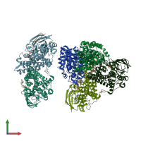
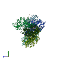
Function and Biology Details
Reaction catalysed:
Glutaryl-CoA + acceptor = (E)-glutaconyl-CoA + reduced acceptor
Biochemical function:
Biological process:
Cellular component:
- not assigned
Sequence domains:
- Acyl-CoA dehydrogenase/oxidase, C-terminal
- Acyl-CoA dehydrogenase-like, C-terminal
- Acyl-CoA dehydrogenase, conserved site
- Acyl-CoA oxidase/dehydrogenase, middle domain
- Acyl-CoA oxidase/dehydrogenase, middle domain superfamily
- Acyl-CoA dehydrogenase/oxidase, N-terminal
- Acyl-CoA dehydrogenase/oxidase, N-terminal domain superfamily
- Acyl-CoA dehydrogenase/oxidase, N-terminal and middle domain superfamily
Structure analysis Details
Assemblies composition:
Assembly name:
Glutaryl-CoA dehydrogenase and peptide (preferred)
PDBe Complex ID:
PDB-CPX-111226 (preferred)
Entry contents:
2 distinct polypeptide molecules
Macromolecules (2 distinct):


