


Function and Biology Details
Reactions catalysed:
RNA(n) + a ribonucleoside 5'-triphosphate = RNA(n+1) + diphosphate.
Hydrolysis of four peptide bonds in the viral precursor polyprotein,commonly with Asp or Glu in the P6 position, Cys or Thr in P1 and Ser orAla in P1'.
a ribonucleoside 5'-triphosphate + H2O = a ribonucleoside 5'-diphosphate+ phosphate + H(+).
ATP + H2O = ADP + phosphate + H(+).
Biochemical function:
Biological process:
Cellular component:
Sequence domains:
- Immunoglobulin C1-set
- MHC class I alpha chain, alpha1 alpha2 domains
- Immunoglobulin/major histocompatibility complex, conserved site
- Immunoglobulin-like domain superfamily
- Immunoglobulin-like domain
- Immunoglobulin-like fold
- Major Histocompatibility Complex/Immunoglobulin
- Beta-2-Microglobulin
4 more domains
Structure analysis Details
Assembly composition:
hetero trimer (preferred)
PDBe Complex ID:
PDB-CPX-137803 (preferred)
Entry contents:
3 distinct polypeptide molecules
Macromolecules (3 distinct):
HLA class I histocompatibility antigen, A alpha chain
Molecule details ›
Chain: A
Length: 293 amino acids
Theoretical weight: 34.01 KDa
Source organism: Homo sapiens
Expression system: Escherichia coli
UniProt:
Sequence domains:
Structure domains:
Length: 293 amino acids
Theoretical weight: 34.01 KDa
Source organism: Homo sapiens
Expression system: Escherichia coli
UniProt:
- Canonical:
 P04439 (Residues: 25-304; Coverage: 81%)
Sequence domains:
Structure domains:
Beta-2-microglobulin
Molecule details ›
Chain: B
Length: 100 amino acids
Theoretical weight: 11.88 KDa
Source organism: Homo sapiens
Expression system: Escherichia coli
UniProt:
Sequence domains: Immunoglobulin C1-set domain
Structure domains: Immunoglobulins
Length: 100 amino acids
Theoretical weight: 11.88 KDa
Source organism: Homo sapiens
Expression system: Escherichia coli
UniProt:
- Canonical: P61769 (Residues: 21-119; Coverage: 100%)
Sequence domains: Immunoglobulin C1-set domain
Structure domains: Immunoglobulins
Serine protease/helicase NS3
Ligands and Environments
No bound ligands
No modified residues
Experiments and Validation Details
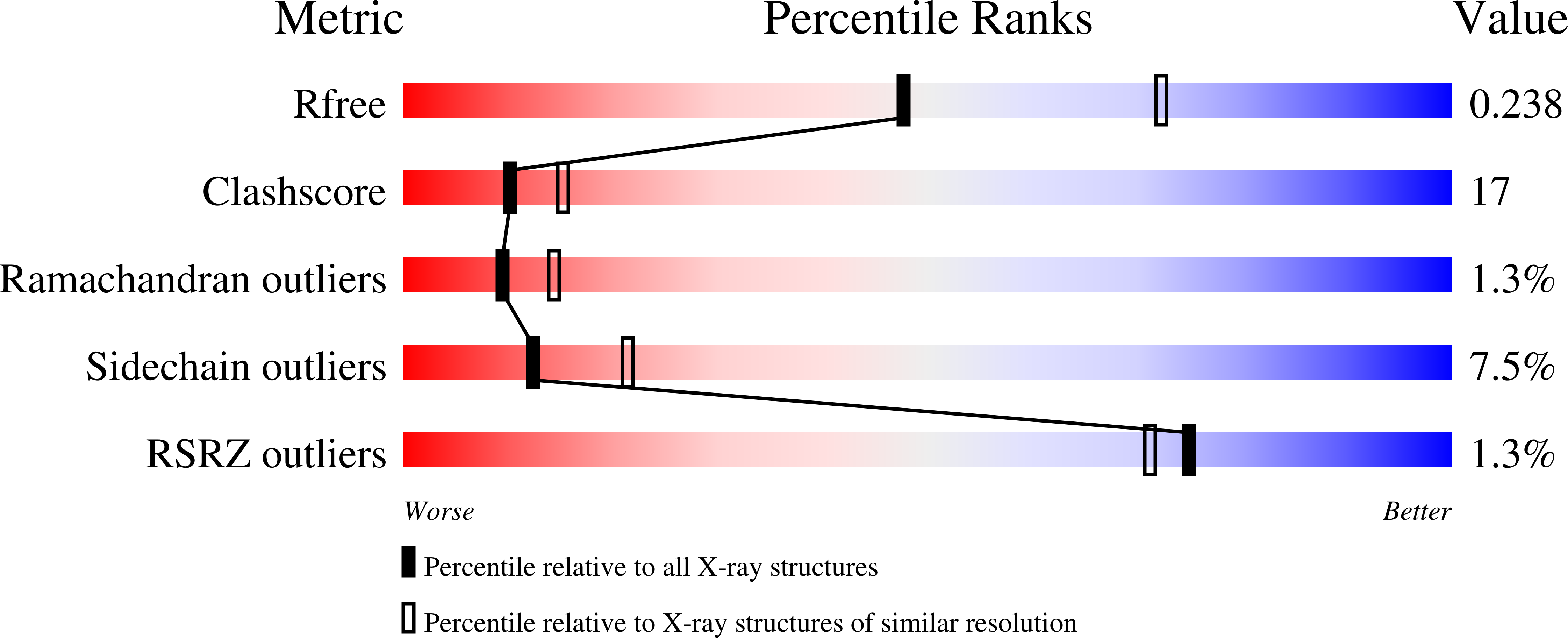
wwPDB Validation report is not available for this entry.
X-ray source:
ESRF BEAMLINE ID29
Spacegroup:
P21
Expression systems:
- Escherichia coli
- Not provided
