


Function and Biology Details
Biochemical function:
- not assigned
Biological process:
- not assigned
Cellular component:
Structure analysis Details
Assembly composition:
monomeric (preferred)
Assembly name:
Kinesin light chain 1 (preferred)
PDBe Complex ID:
PDB-CPX-170551 (preferred)
Entry contents:
1 distinct polypeptide molecule
Macromolecule:
Kinesin light chain 1
Molecule details ›
Chain: A
Length: 311 amino acids
Theoretical weight: 35.44 KDa
Source organism: Homo sapiens
Expression system: Escherichia coli
UniProt:
Sequence domains:
Structure domains: Tetratricopeptide repeat domain
Length: 311 amino acids
Theoretical weight: 35.44 KDa
Source organism: Homo sapiens
Expression system: Escherichia coli
UniProt:
- Canonical:
 Q07866 (Residues: 203-497; Coverage: 52%)
Sequence domains:
Structure domains: Tetratricopeptide repeat domain
Ligands and Environments
No bound ligands
No modified residues
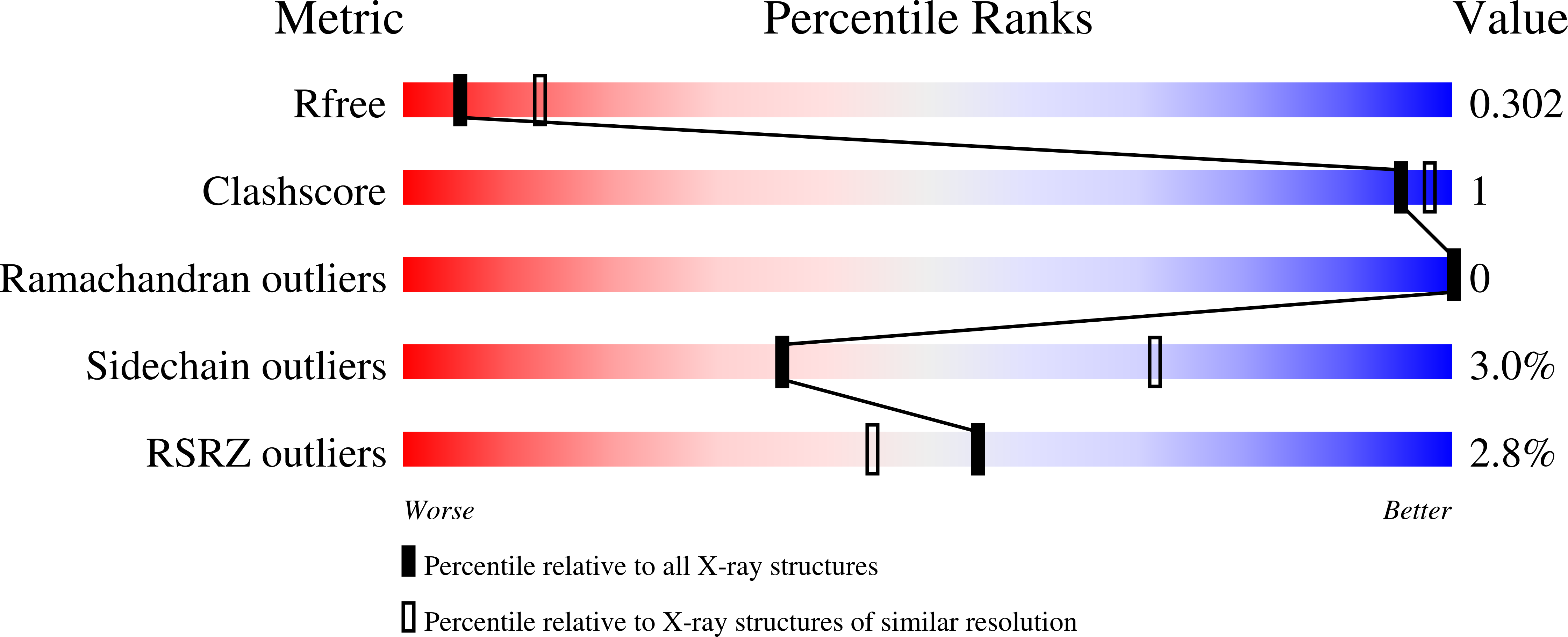
Experiments and Validation Details
wwPDB Validation report is not available for this entry.
X-ray source:
CLSI BEAMLINE 08ID-1
Spacegroup:
P3121
Expression system: Escherichia coli
