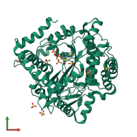
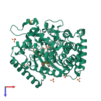
3nzt

X-ray diffraction
2Å resolution

2.0 Angstrom Crystal structure of Glutamate--Cysteine Ligase (gshA) ftom Francisella tularensis in Complex with AMP

Released:
Entry authors: Minasov G, Halavaty A, Shuvalova L, Dubrovska I, Winsor J, Papazisi L, Anderson WF, Center for Structural Genomics of Infectious Diseases (CSGID)

Function and Biology Details

Reaction catalysed:
ATP + L-glutamate + L-cysteine = ADP + phosphate + gamma-L-glutamyl-L-cysteine
Biochemical function:
Cellular component:

Structure analysis Details

Assembly composition:
monomeric (preferred)
PDBe Complex ID:
PDB-CPX-177709 (preferred)
Entry contents:
1 distinct polypeptide molecule
Macromolecule:
Glutamate--cysteine ligase Chain: A
Molecule details ›
Chain: A
Length: 525 amino acids
Theoretical weight: 59.72 KDa
Source organism: Francisella tularensis subsp. tularensis
Expression system: Escherichia coli BL21(DE3)
UniProt:
  • Canonical: Q5NHS8 (Residues: 1-501; Coverage: 100%)
Gene names: FTT_0367c, gshA
Sequence domains: Glutamate-cysteine ligase
Structure domains: Creatine Kinase; Chain A, domain 2

Ligands and Environments

2 bound ligands:
No modified residues

Experiments and Validation Details

Entry percentile scores
X-ray source: APS BEAMLINE 21-ID-F
Spacegroup: P65
Unit cell:
a: 89.849Å b: 89.849Å c: 151.468Å
α: 90° β: 90° γ: 120°
R-values:
R R work R free
0.165 0.164 0.196
Expression system: Escherichia coli BL21(DE3)