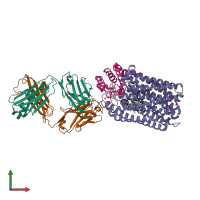
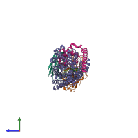
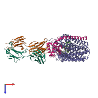
3o0r

X-ray diffraction
2.7Å resolution

Crystal structure of nitric oxide reductase from Pseudomonas aeruginosa in complex with antibody fragment

Released:

Function and Biology Details

Reaction catalysed:
Nitrous oxide + 2 ferricytochrome c + H(2)O = 2 nitric oxide + 2 ferrocytochrome c + 2 H(+)
Biochemical function:
Cellular component:

Structure analysis Details

Assembly composition:
hetero tetramer (preferred)
PDBe Complex ID:
PDB-CPX-212293 (preferred)
Entry contents:
4 distinct polypeptide molecules
Macromolecules (4 distinct):
antibody fab fragment light chain Chain: L
Molecule details ›
Chain: L
Length: 213 amino acids
Theoretical weight: 23.74 KDa
Source organism: Mus musculus
Structure domains: Immunoglobulins
antibody fab fragment heavy chain Chain: H
Molecule details ›
Chain: H
Length: 225 amino acids
Theoretical weight: 24.09 KDa
Source organism: Mus musculus
Structure domains: Immunoglobulins
Nitric oxide reductase subunit B Chain: B
Molecule details ›
Chain: B
Length: 465 amino acids
Theoretical weight: 52.22 KDa
Source organism: Pseudomonas aeruginosa PAO1
UniProt:
  • Canonical: Q59647 (Residues: 1-466; Coverage: 100%)
Gene names: PA0524, norB
Sequence domains: Cytochrome C and Quinol oxidase polypeptide I
Structure domains: Cytochrome c oxidase-like, subunit I domain
Nitric oxide reductase subunit C Chain: C
Molecule details ›
Chain: C
Length: 146 amino acids
Theoretical weight: 16.37 KDa
Source organism: Pseudomonas aeruginosa PAO1
UniProt:
  • Canonical: Q59646 (Residues: 1-146; Coverage: 100%)
Gene names: PA0523, norC
Sequence domains: Cytochrome c
Structure domains: Cytochrome c-like domain

Ligands and Environments


Cofactor: Ligand HEM 2 x HEM

Cofactor: Ligand HEC 1 x HEC
3 bound ligands:
No modified residues

Experiments and Validation Details

Entry percentile scores
X-ray source: SPRING-8 BEAMLINE BL41XU, SPRING-8 BEAMLINE BL26B2
Spacegroup: P212121
Unit cell:
a: 90.47Å b: 104.52Å c: 195.36Å
α: 90° β: 90° γ: 90°
R-values:
R R work R free
0.188 0.185 0.247