Function and Biology Details
Reaction catalysed:
Limited hydrolysis of proteins of the neuroexocytosis apparatus, synaptobrevin (also known as neuronal vesicle-associated membrane protein, VAMP), synaptosome-associated protein of 25 kDa (SNAP25) or syntaxin. No detected action on small molecule substrates.
Biochemical function:
- not assigned
Biological process:
- not assigned
Cellular component:
Structure analysis Details
Assembly composition:
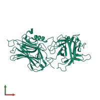
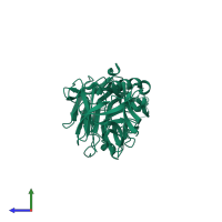
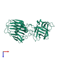
monomeric (preferred)
Assembly name:
Botulinum neurotoxin D light chain (preferred)
PDBe Complex ID:
PDB-CPX-148553 (preferred)
Entry contents:
1 distinct polypeptide molecule
Macromolecule:
Ligands and Environments
No bound ligands
No modified residues
Experiments and Validation Details
wwPDB Validation report is not available for this entry.
X-ray source:
NSLS BEAMLINE X29A
Spacegroup:
P212121
Expression system: Escherichia coli


