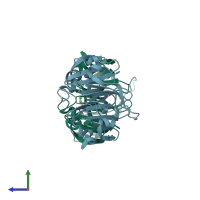
3oyo

X-ray diffraction
2.1Å resolution

Crystal structure of hemopexin fold protein CP4 from cow pea

Released:
Source organism: Vigna unguiculata
Primary publication:
The structure of a haemopexin-fold protein from cow pea (Vigna unguiculata) suggests functional diversity of haemopexins in plants.
Acta Crystallogr Sect F Struct Biol Cryst Commun 67 193-200 (2011)
PMID: 21301085

Function and Biology Details

Biochemical function:
Biological process:
  • not assigned
Cellular component:
  • not assigned

Structure analysis Details

Assembly composition:
monomeric (preferred)
PDBe Complex ID:
PDB-CPX-123446 (preferred)
Entry contents:
1 distinct polypeptide molecule
Macromolecule:
Hemopexin fold protein CP4 Chains: A, B
Molecule details ›
Chains: A, B
Length: 225 amino acids
Theoretical weight: 24.74 KDa
Source organism: Vigna unguiculata
UniProt:
  • Canonical: F2Z290 (Residues: 1-225; Coverage: 100%)
Sequence domains: Hemopexin
Structure domains: Hemopexin-like domain

Ligands and Environments

3 bound ligands:
No modified residues

Experiments and Validation Details

Entry percentile scores
X-ray source: RIGAKU RU300
Spacegroup: C2
Unit cell:
a: 125.087Å b: 60.138Å c: 67.574Å
α: 90° β: 111.18° γ: 90°
R-values:
R R work R free
0.209 0.205 0.246