3pei

X-ray diffraction
2.7Å resolution

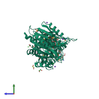
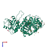
Crystal Structure of Cytosol Aminopeptidase from Francisella tularensis

Released:
Entry authors: Maltseva N, Kim Y, Gu M, Anderson WF, Joachimiak A, Center for Structural Genomics of Infectious Diseases (CSGID)

Function and Biology Details

Reactions catalysed:
Release of an N-terminal amino acid, Xaa-|-Yaa-, in which Xaa is preferably Leu, but may be other amino acids including Pro although not Arg or Lys, and Yaa may be Pro. Amino acid amides and methyl esters are also readily hydrolyzed, but rates on arylamides are exceedingly low.
Release of an N-terminal amino acid, preferentially leucine, but not glutamic or aspartic acids.
Biochemical function:
Biological process:
Cellular component:

Structure analysis Details

Assembly composition:
homo hexamer (preferred)
PDBe Complex ID:
PDB-CPX-177661 (preferred)
Entry contents:
1 distinct polypeptide molecule
Macromolecule:
Probable cytosol aminopeptidase Chain: A
Molecule details ›
Chain: A
Length: 486 amino acids
Theoretical weight: 53.95 KDa
Source organism: Francisella tularensis subsp. tularensis SCHU S4
Expression system: Escherichia coli BL21
UniProt:
  • Canonical: Q5NFC1 (Residues: 1-479; Coverage: 100%)
Gene names: FTT_1318c, pepA
Sequence domains:
Structure domains:

Ligands and Environments

1 modified residue:

Experiments and Validation Details

Entry percentile scores
X-ray source: APS BEAMLINE 19-ID
Spacegroup: P6322
Unit cell:
a: 161.483Å b: 161.483Å c: 105.27Å
α: 90° β: 90° γ: 120°
R-values:
R R work R free
0.167 0.165 0.21
Expression system: Escherichia coli BL21