3pox

X-ray diffraction
2Å resolution

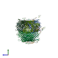
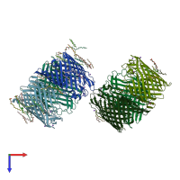
Crystal Structure of E.coli OmpF porin in lipidic cubic phase: space group P1

Released:
Source organism: Escherichia coli BL21
Primary publication:
Structure of Escherichia coli OmpF porin from lipidic mesophase.
J Struct Biol 178 311-8 (2012)
PMID: 22484237

Function and Biology Details

Structure analysis Details

Assembly composition:
homo trimer (preferred)
Assembly name:
PDBe Complex ID:
PDB-CPX-136399 (preferred)
Entry contents:
1 distinct polypeptide molecule
Macromolecule:
Outer membrane porin F Chains: A, B, C, D, E, F
Molecule details ›
Chains: A, B, C, D, E, F
Length: 340 amino acids
Theoretical weight: 37.11 KDa
Source organism: Escherichia coli BL21
UniProt:
  • Canonical: P02931 (Residues: 23-362; Coverage: 100%)
Gene names: JW0912, b0929, cmlB, coa, cry, ompF, tolF
Sequence domains: Gram-negative porin
Structure domains: Porin

Ligands and Environments

3 bound ligands:
No modified residues

Experiments and Validation Details

Entry percentile scores
X-ray source: DIAMOND BEAMLINE I24
Spacegroup: P1
Unit cell:
a: 79.574Å b: 86.017Å c: 116.558Å
α: 83.28° β: 78.46° γ: 89.89°
R-values:
R R work R free
0.19 0.188 0.225