


Function and Biology Details
Reaction catalysed:
Peptidylproline (omega=180) = peptidylproline (omega=0)
Biochemical function:
Biological process:
- not assigned
Cellular component:
- not assigned
Structure analysis Details
Assembly composition:
homo dimer (preferred)
Assembly name:
Long-type peptidyl-prolyl cis-trans isomerase (preferred)
PDBe Complex ID:
PDB-CPX-176588 (preferred)
Entry contents:
1 distinct polypeptide molecule
Macromolecule:
Long-type peptidyl-prolyl cis-trans isomerase
Molecule details ›
Chains: A, B
Length: 157 amino acids
Theoretical weight: 17.51 KDa
Source organism: Methanocaldococcus jannaschii
Expression system: Escherichia coli
UniProt:
Sequence domains:
Structure domains: Chitinase A; domain 3
Length: 157 amino acids
Theoretical weight: 17.51 KDa
Source organism: Methanocaldococcus jannaschii
Expression system: Escherichia coli
UniProt:
- Canonical:
 Q58235 (Residues: 1-150; Coverage: 65%)
Sequence domains:
Structure domains: Chitinase A; domain 3
Ligands and Environments
No bound ligands
No modified residues
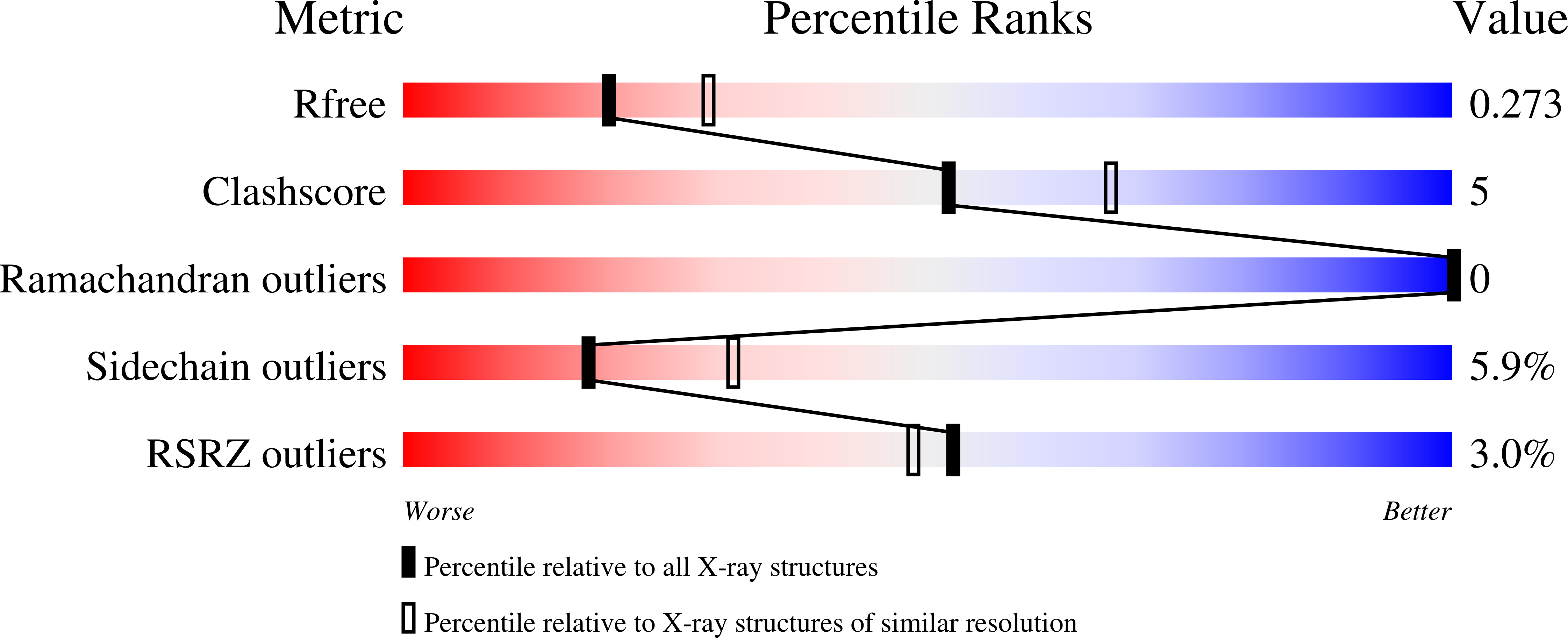
Experiments and Validation Details
wwPDB Validation report is not available for this entry.
X-ray source:
NSLS BEAMLINE X4A
Spacegroup:
P21
Expression system: Escherichia coli
