3ree

X-ray diffraction
1.76Å resolution

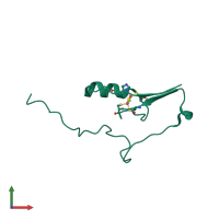
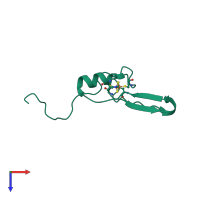
Crystal structure of mitoNEET

Released:

Function and Biology Details

Reaction catalysed:
L-cysteine + 2-oxoglutarate = 2-oxo-3-sulfanylpropanoate + L-glutamate
Biochemical function:
Biological process:

Structure analysis Details

Assembly composition:
homo dimer (preferred)
PDBe Complex ID:
PDB-CPX-192731 (preferred)
Entry contents:
1 distinct polypeptide molecule
Macromolecule:
CDGSH iron-sulfur domain-containing protein 1 Chain: A
Molecule details ›
Chain: A
Length: 84 amino acids
Theoretical weight: 9.78 KDa
Source organism: Homo sapiens
Expression system: Escherichia coli
UniProt:
  • Canonical: Q9NZ45 (Residues: 32-108; Coverage: 71%)
Gene names: C10orf70, CISD1, MDS029, ZCD1
Sequence domains: Iron-binding zinc finger CDGSH type
Structure domains: CDGSH iron-sulfur domain, mitoNEET-type

Ligands and Environments

1 bound ligand:
No modified residues

Experiments and Validation Details

Entry percentile scores
X-ray source: APS BEAMLINE 21-ID-F
Spacegroup: I4122
Unit cell:
a: 58.64Å b: 58.64Å c: 177.61Å
α: 90° β: 90° γ: 90°
R-values:
R R work R free
0.203 0.202 0.228
Expression system: Escherichia coli