3rty

X-ray diffraction
2.85Å resolution

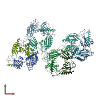
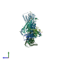
Structure of an Enclosed Dimer Formed by The Drosophila Period Protein

Released:
Source organism: Drosophila melanogaster
Primary publication:
Structure of an enclosed dimer formed by the Drosophila period protein.
J Mol Biol 413 561-72 (2011)
PMID: 21907720

Function and Biology Details

Biochemical function:
  • not assigned
Cellular component:
  • not assigned

Structure analysis Details

Assembly composition:
homo dimer (preferred)
PDBe Complex ID:
PDB-CPX-139687 (preferred)
Entry contents:
1 distinct polypeptide molecule
Macromolecule:
Period circadian protein Chains: A, B, C, D, E, F, G, H
Molecule details ›
Chains: A, B, C, D, E, F, G, H
Length: 339 amino acids
Theoretical weight: 38.19 KDa
Source organism: Drosophila melanogaster
Expression system: Escherichia coli
UniProt:
  • Canonical: P07663 (Residues: 236-574; Coverage: 28%)
Gene names: CG2647, per
Sequence domains:
Structure domains: PAS domain

Ligands and Environments

1 bound ligand:
No modified residues

Experiments and Validation Details

Entry percentile scores
X-ray source: ALS BEAMLINE 8.2.1
Spacegroup: P1
Unit cell:
a: 60.414Å b: 94.697Å c: 141.03Å
α: 88.19° β: 89.63° γ: 89.87°
R-values:
R R work R free
0.239 0.239 0.289
Expression system: Escherichia coli