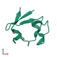
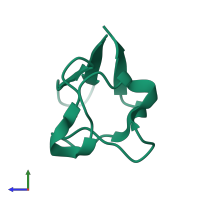
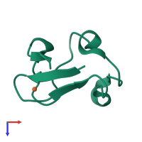
3ryg

Neutron Diffraction
1.75Å resolution

128 hours neutron structure of perdeuterated rubredoxin

Released:
Source organism: Pyrococcus furiosus DSM 3638
Primary publication:
Rapid visualization of hydrogen positions in protein neutron crystallographic structures.
Acta Crystallogr D Biol Crystallogr 68 35-41 (2012)
PMID: 22194331

Function and Biology Details

Biochemical function:
Biological process:
Cellular component:
  • not assigned

Structure analysis Details

Assembly composition:
monomeric (preferred)
Assembly name:
PDBe Complex ID:
PDB-CPX-150200 (preferred)
Entry contents:
1 distinct polypeptide molecule
Macromolecule:
Rubredoxin Chain: A
Molecule details ›
Chain: A
Length: 54 amino acids
Theoretical weight: 6.03 KDa
Source organism: Pyrococcus furiosus DSM 3638
Expression system: Escherichia coli BL21(DE3)
UniProt:
  • Canonical: P24297 (Residues: 1-54; Coverage: 100%)
Gene names: PF1282, rub
Sequence domains: Rubredoxin
Structure domains: Rubrerythrin, domain 2

Ligands and Environments

2 bound ligands:
No modified residues

Experiments and Validation Details

Spacegroup: P212121
Unit cell:
a: 34.37Å b: 35.34Å c: 44.11Å α: 90° β: 90° γ: 90°
R-values:
R R work R free 0.182 0.181 0.2
Expression system: Escherichia coli BL21(DE3)