


Function and Biology Details
Reaction catalysed:
Protein tyrosine phosphate + H(2)O = protein tyrosine + phosphate
Biochemical function:
Biological process:
Cellular component:
- not assigned
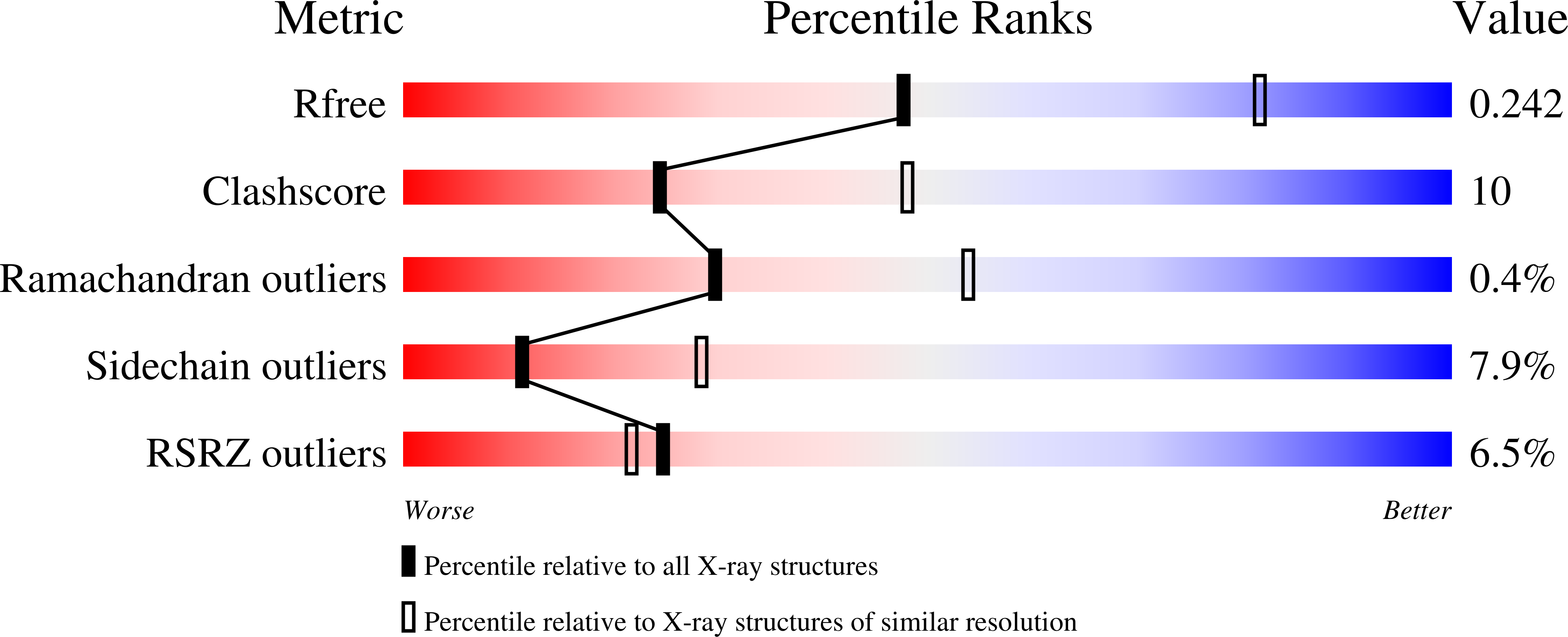
Structure analysis Details
Assembly composition:
monomeric (preferred)
Assembly name:
Tyrosine-protein phosphatase 10D (preferred)
PDBe Complex ID:
PDB-CPX-153096 (preferred)
Entry contents:
1 distinct polypeptide molecule
Macromolecule:
Tyrosine-protein phosphatase 10D
Molecule details ›
Chains: A, B
Length: 307 amino acids
Theoretical weight: 35.95 KDa
Source organism: Drosophila melanogaster
Expression system: Escherichia coli
UniProt:
Sequence domains: Protein-tyrosine phosphatase
Structure domains: Protein tyrosine phosphatase superfamily
Length: 307 amino acids
Theoretical weight: 35.95 KDa
Source organism: Drosophila melanogaster
Expression system: Escherichia coli
UniProt:
- Canonical:
 P35992 (Residues: 1250-1533; Coverage: 15%)
Sequence domains: Protein-tyrosine phosphatase
Structure domains: Protein tyrosine phosphatase superfamily


