


Function and Biology Details
Reaction catalysed:
Hydrolysis of alpha-(2->3)-, alpha-(2->6)-, alpha-(2->8)- glycosidic linkages of terminal sialic acid residues in oligosaccharides, glycoproteins, glycolipids, colominic acid and synthetic substrates.
Biochemical function:
Biological process:
Cellular component:
Structure analysis Details
Assembly composition:
homo octamer (preferred)
Assembly name:
Hemagglutinin-neuraminidase (preferred)
PDBe Complex ID:
PDB-CPX-146264 (preferred)
Entry contents:
1 distinct polypeptide molecule
Macromolecule:
Hemagglutinin-neuraminidase
Molecule details ›
Chains: A, B, E, F
Length: 537 amino acids
Theoretical weight: 58.7 KDa
Source organism: Newcastle disease virus (STRAIN AUSTRALIA-VICTORIA/32)
Expression system: Spodoptera frugiperda
UniProt:
Sequence domains: Haemagglutinin-neuraminidase
Structure domains: Neuraminidase
Length: 537 amino acids
Theoretical weight: 58.7 KDa
Source organism: Newcastle disease virus (STRAIN AUSTRALIA-VICTORIA/32)
Expression system: Spodoptera frugiperda
UniProt:
- Canonical:
 P12554 (Residues: 49-570; Coverage: 92%)
Sequence domains: Haemagglutinin-neuraminidase
Structure domains: Neuraminidase
Ligands and Environments
No bound ligands
No modified residues
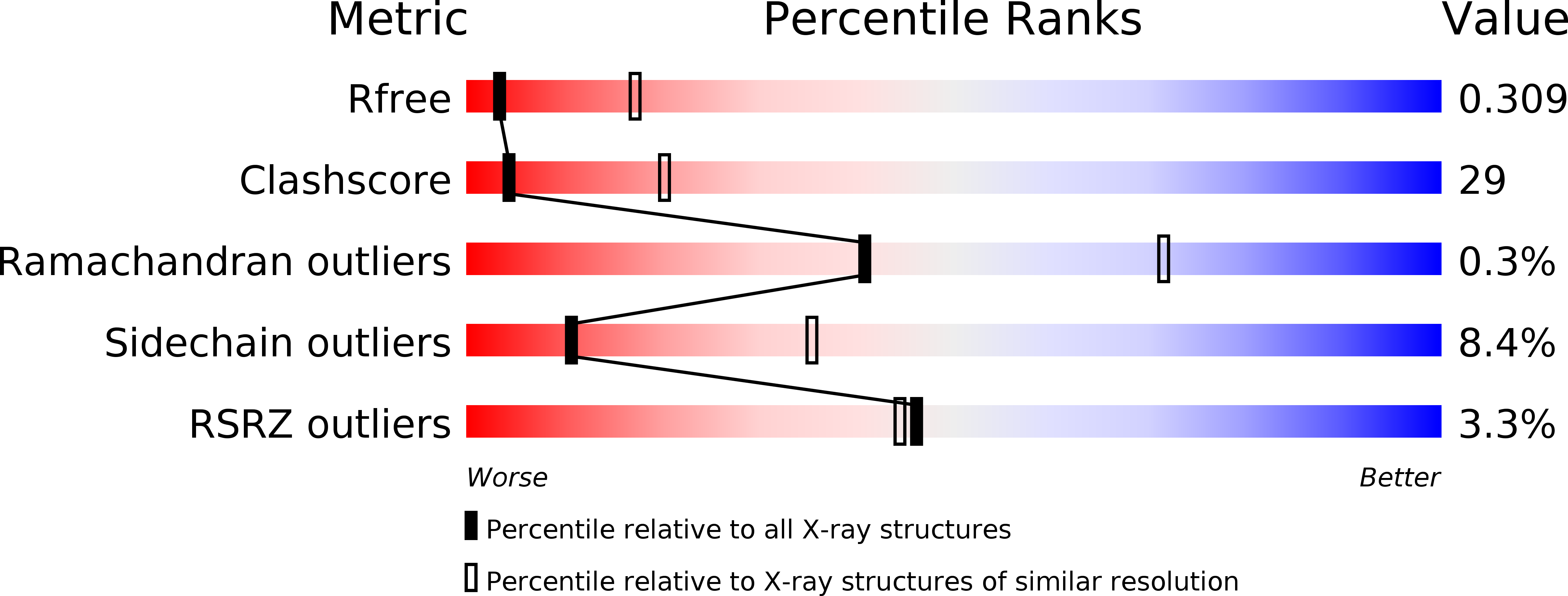
Experiments and Validation Details

X-ray source:
APS BEAMLINE 21-ID-D
Spacegroup:
P43212
Expression system: Spodoptera frugiperda