


Function and Biology Details
Biochemical function:
Biological process:
- not assigned
Cellular component:
- not assigned
Structure analysis Details
Assembly composition:
homo dimer (preferred)
Assembly name:
Stromal interaction molecule 1 (preferred)
PDBe Complex ID:
PDB-CPX-124516 (preferred)
Entry contents:
1 distinct polypeptide molecule
Macromolecule:
Stromal interaction molecule 1
Molecule details ›
Chains: A, B
Length: 136 amino acids
Theoretical weight: 15.62 KDa
Source organism: Caenorhabditis elegans
Expression system: Escherichia coli
UniProt:
Sequence domains: STIM1 Orai1-activating region
Structure domains: Helix Hairpins
Length: 136 amino acids
Theoretical weight: 15.62 KDa
Source organism: Caenorhabditis elegans
Expression system: Escherichia coli
UniProt:
- Canonical:
 G5EF60 (Residues: 256-391; Coverage: 27%)
Sequence domains: STIM1 Orai1-activating region
Structure domains: Helix Hairpins
Ligands and Environments
No bound ligands
No modified residues
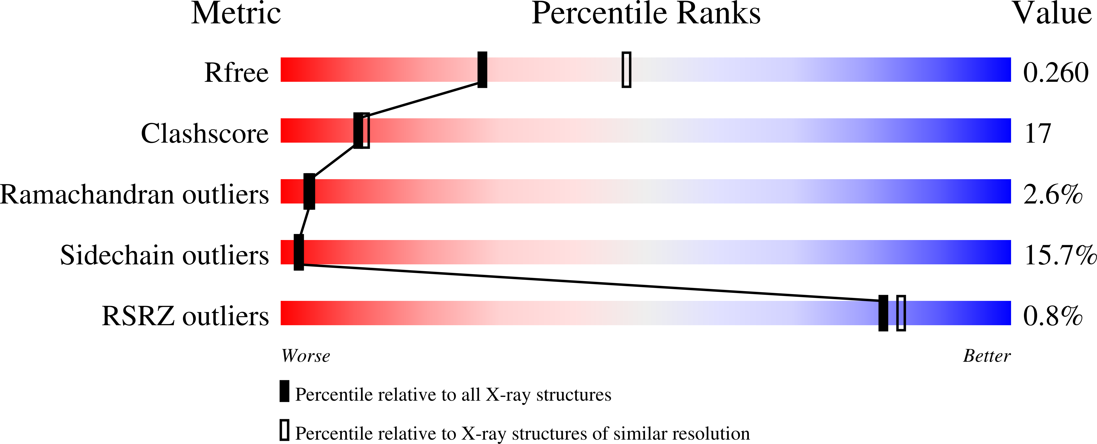
Experiments and Validation Details
wwPDB Validation report is not available for this entry.
X-ray source:
SSRF BEAMLINE BL17U
Spacegroup:
P32
Expression system: Escherichia coli
