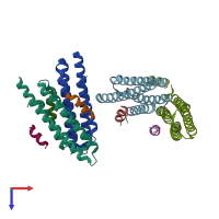
3u3f

X-ray diffraction
3.1Å resolution

Structural basis for the interaction of Pyk2 PAT domain with paxillin LD motifs

Released:

Function and Biology Details

Reaction catalysed:
ATP + a [protein]-L-tyrosine = ADP + a [protein]-L-tyrosine phosphate
Biochemical function:
Biological process:
Cellular component:

Structure analysis Details

Assemblies composition:
hetero trimer (preferred)
hetero dimer
PDBe Complex ID:
PDB-CPX-155840 (preferred)
Entry contents:
2 distinct polypeptide molecules
Macromolecules (2 distinct):
Protein-tyrosine kinase 2-beta Chains: A, B, C, D
Molecule details ›
Chains: A, B, C, D
Length: 139 amino acids
Theoretical weight: 15.26 KDa
Source organism: Homo sapiens
Expression system: Escherichia coli BL21(DE3)
UniProt:
  • Canonical: Q14289 (Residues: 871-1005; Coverage: 13%)
Gene names: FAK2, PTK2B, PYK2, RAFTK
Sequence domains: Focal adhesion targeting region
Structure domains: Nucleotidyltransferases domain 2
Paxillin Chains: E, F, G, H, I, J
Molecule details ›
Chains: E, F, G, H, I, J
Length: 17 amino acids
Theoretical weight: 1.92 KDa
Source organism: Homo sapiens
Expression system: Not provided
UniProt:
  • Canonical: P49023 (Residues: 261-277; Coverage: 3%)
Gene name: PXN

Ligands and Environments

No bound ligands
No modified residues

Experiments and Validation Details

Entry percentile scores
X-ray source: APS BEAMLINE 22-BM
Spacegroup: P212121
Unit cell:
a: 83.599Å b: 83.747Å c: 170.545Å
α: 90° β: 90° γ: 90°
R-values:
R R work R free
0.22 0.218 0.266
Expression systems:
  • Escherichia coli BL21(DE3)
  • Not provided