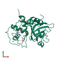
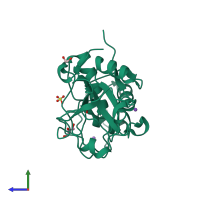
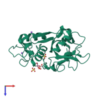
3u8e

X-ray diffraction
1.31Å resolution

Crystal Structure of Cysteine Protease from Bulbs of Crocus sativus at 1.3 A Resolution

Released:
Source organism: Crocus sativus
Entry authors: Iqbal S, Akrem A, Buck F, Perbandt M, Banumathi S, Betzel C

Function and Biology Details

Structure analysis Details

Assembly composition:
homo trimer (preferred)
PDBe Complex ID:
PDB-CPX-124614 (preferred)
Entry contents:
1 distinct polypeptide molecule
Macromolecule:
Peptidase C1A papain C-terminal domain-containing protein Chain: A
Molecule details ›
Chain: A
Length: 222 amino acids
Theoretical weight: 23.21 KDa
Source organism: Crocus sativus
UniProt:
  • Canonical: G8JLU6 (Residues: 1-222; Coverage: 100%)
Sequence domains: Papain family cysteine protease
Structure domains: Cysteine proteinases

Ligands and Environments

3 bound ligands:
No modified residues

Experiments and Validation Details

Entry percentile scores
X-ray source: ALS BEAMLINE 8.3.1
Spacegroup: R3
Unit cell:
a: 97.97Å b: 97.97Å c: 48.78Å
α: 90° β: 90° γ: 120°
R-values:
R R work R free
0.189 0.144 0.189