3ub5

X-ray diffraction
2.2Å resolution

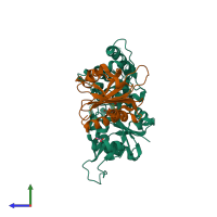
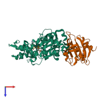
Profilin:actin with a wide open nucleotide cleft

Released:
Source organism: Bos taurus
Primary publication:
Structural basis for profilin-mediated actin nucleotide exchange.
J Mol Biol 418 103-16 (2012)
PMID: 22366544

Function and Biology Details

Structure analysis Details

Assembly composition:
hetero dimer (preferred)
PDBe Complex ID:
PDB-CPX-135936 (preferred)
Entry contents:
2 distinct polypeptide molecules
Macromolecules (2 distinct):
Actin, cytoplasmic 1 Chain: A
Molecule details ›
Chain: A
Length: 374 amino acids
Theoretical weight: 41.65 KDa
Source organism: Bos taurus
UniProt:
  • Canonical: P60712 (Residues: 2-375; Coverage: 100%)
Gene name: ACTB
Sequence domains: Actin
Structure domains:
Profilin-1 Chain: P
Molecule details ›
Chain: P
Length: 140 amino acids
Theoretical weight: 14.97 KDa
Source organism: Bos taurus
UniProt:
  • Canonical: P02584 (Residues: 2-140; Coverage: 99%)
Gene name: PFN1
Sequence domains: Profilin
Structure domains: Dynein light chain 2a, cytoplasmic

Ligands and Environments

No modified residues

Experiments and Validation Details

Entry percentile scores
X-ray source: RIGAKU FR-E SUPERBRIGHT
Spacegroup: P212121
Unit cell:
a: 38Å b: 72Å c: 186.8Å
α: 90° β: 90° γ: 90°
R-values:
R R work R free
0.219 0.215 0.29