


Function and Biology Details
Biochemical function:
- not assigned
Biological process:
Cellular component:
- not assigned
Structure analysis Details
Assemblies composition:
Assembly name:
PDBe Complex ID:
PDB-CPX-154134 (preferred)
Entry contents:
2 distinct polypeptide molecules
Macromolecules (2 distinct):
Mitochondrial fission 1 protein
Molecule details ›
Chains: A, C
Length: 129 amino acids
Theoretical weight: 15.13 KDa
Source organism: Saccharomyces cerevisiae S288C
Expression system: Escherichia coli
UniProt:
Sequence domains:
Structure domains: Tetratricopeptide repeat domain
Length: 129 amino acids
Theoretical weight: 15.13 KDa
Source organism: Saccharomyces cerevisiae S288C
Expression system: Escherichia coli
UniProt:
- Canonical:
 P40515 (Residues: 1-129; Coverage: 83%)
Sequence domains:
Structure domains: Tetratricopeptide repeat domain
Mitochondrial division protein 1
Molecule details ›
Chains: B, D
Length: 242 amino acids
Theoretical weight: 27.14 KDa
Source organism: Saccharomyces cerevisiae S288C
Expression system: Escherichia coli
UniProt:
Sequence domains: Mitochondrial division protein 1
Structure domains: Methane Monooxygenase Hydroxylase; Chain G, domain 1
Length: 242 amino acids
Theoretical weight: 27.14 KDa
Source organism: Saccharomyces cerevisiae S288C
Expression system: Escherichia coli
UniProt:
- Canonical: P47025 (Residues: 94-314; Coverage: 31%)
Sequence domains: Mitochondrial division protein 1
Structure domains: Methane Monooxygenase Hydroxylase; Chain G, domain 1
Ligands and Environments
No bound ligands
No modified residues
Experiments and Validation Details
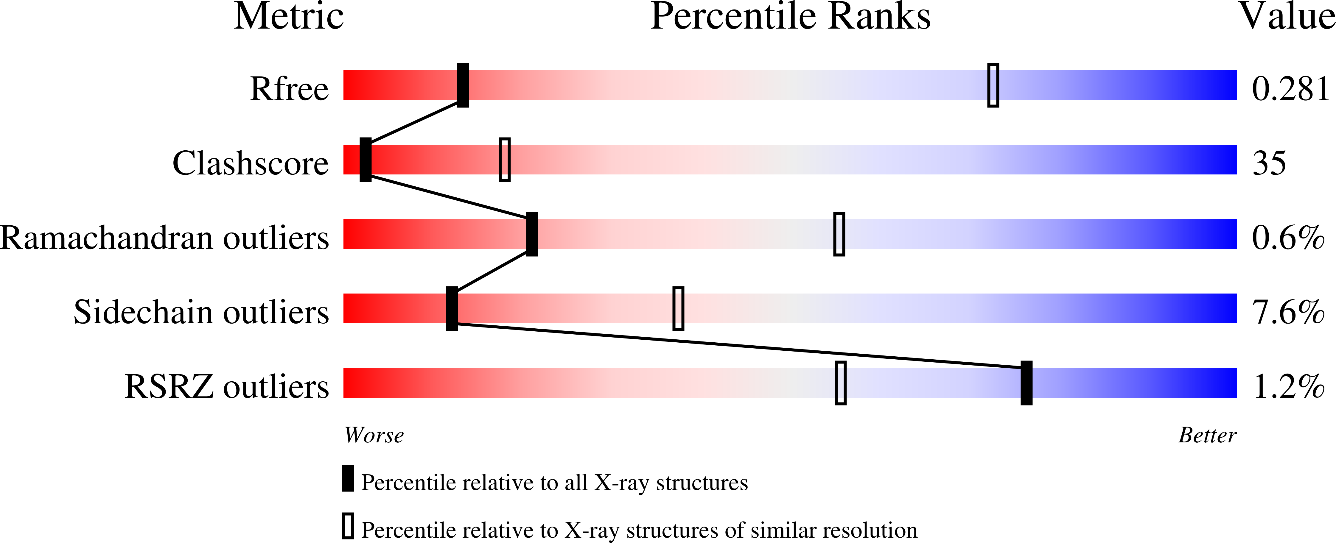
wwPDB Validation report is not available for this entry.
X-ray source:
SSRL BEAMLINE BL9-1
Spacegroup:
I422
Expression system: Escherichia coli
