


Function and Biology Details
Reaction catalysed:
Phosphatidylcholine + H(2)O = 1-acylglycerophosphocholine + a carboxylate
Biochemical function:
Biological process:
Cellular component:
Structure analysis Details
Assemblies composition:
Assembly name:
PDBe Complex ID:
PDB-CPX-143462 (preferred)
Entry contents:
1 distinct polypeptide molecule
Macromolecule:
Neutral phospholipase A2 homolog taipoxin beta chain 2
Molecule details ›
Chains: A, B
Length: 118 amino acids
Theoretical weight: 13.33 KDa
Source organism: Oxyuranus scutellatus scutellatus
UniProt:
Structure domains: Phospholipase A2 domain
Length: 118 amino acids
Theoretical weight: 13.33 KDa
Source organism: Oxyuranus scutellatus scutellatus
UniProt:
- Canonical:
 P0CG57 (Residues: 1-118; Coverage: 100%)
Structure domains: Phospholipase A2 domain
Ligands and Environments
No bound ligands
No modified residues
Experiments and Validation Details
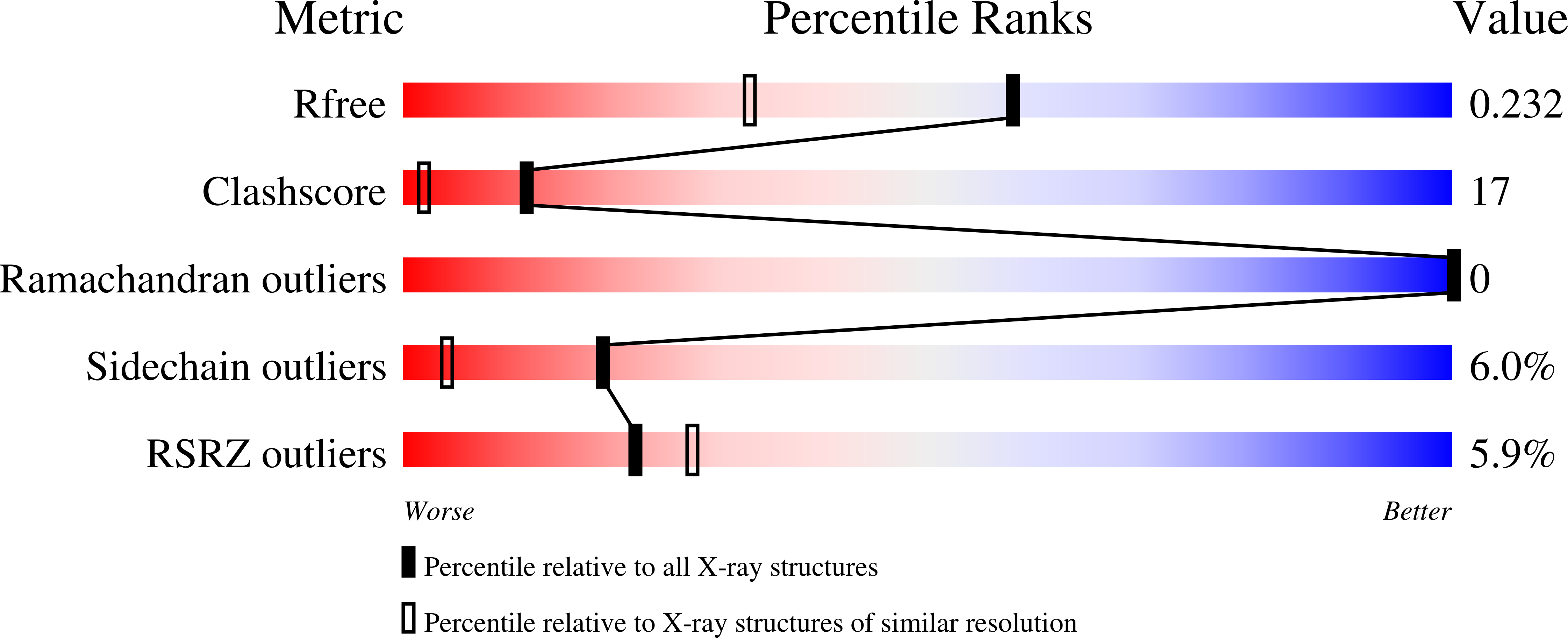
wwPDB Validation report is not available for this entry.
X-ray source:
ESRF BEAMLINE BM14
Spacegroup:
P21
